 Explore
 Validate
 Learn
Learn Western blot
Antibody data
- Antibody Data
- Antigen structure
- References [0]
- Comments [0]
- Validations
- Western blot [3]
- Immunohistochemistry [9]
Submit
Validation data
Reference
Comment
Report error
- Product number
- HPA015083 - Provider product page

- Provider
- Atlas Antibodies
- Proper citation
- Atlas Antibodies Cat#HPA015083, RRID:AB_2669151
- Product name
- Anti-STK10
- Antibody type
- Polyclonal
- Description
- Polyclonal Antibody against Human STK10, Gene description: serine/threonine kinase 10, Alternative Gene Names: LOK, PRO2729, Validated applications: IHC, WB, Uniprot ID: O94804, Storage: Store at +4°C for short term storage. Long time storage is recommended at -20°C.
- Reactivity
- Human
- Host
- Rabbit
- Conjugate
- Unconjugated
- Isotype
- IgG
- Vial size
- 100 µl
- Concentration
- 0.1 mg/ml
- Storage
- Store at +4°C for short term storage. Long time storage is recommended at -20°C.
No comments: Submit comment
Supportive validation
Enhanced validation
Supportive validation
- Submitted by
- Atlas Antibodies (provider)
- Enhanced method
- Orthogonal validation
- Main image

- Experimental details
- Western blot analysis in human cell lines SK-MEL-30 and HeLa using Anti-STK10 antibody. Corresponding STK10 RNA-seq data are presented for the same cell lines. Loading control: Anti-COX4I1.

Enhanced validation
- Submitted by
- Atlas Antibodies (provider)
- Enhanced method
- Orthogonal validation
- Main image

- Experimental details
- Western blot analysis in human cell lines SK-MEL-30 and HeLa using Anti-STK10 antibody. Corresponding STK10 RNA-seq data are presented for the same cell lines. Loading control: Anti-COX4I1.
- Sample type
- Human
- Protocol
- Protocol

Supportive validation
- Submitted by
- Atlas Antibodies (provider)
- Main image

- Experimental details
- Lane 1: Marker [kDa] 230, 130, 95, 72, 56, 36, 28, 17, 11Lane 2: Human cell line RT-4

Enhanced validation
Enhanced validation
Supportive validation
- Submitted by
- Atlas Antibodies (provider)
- Enhanced method
- Orthogonal validation
- Main image
- Experimental details
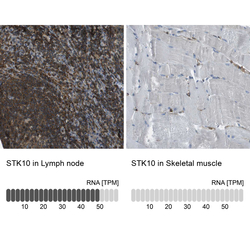
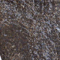
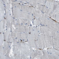
- Immunohistochemistry analysis in human lymph node and skeletal muscle tissues using Anti-STK10 antibody. Corresponding STK10 RNA-seq data are presented for the same tissues.
- Sample type
- HUMAN

Enhanced validation
- Submitted by
- Atlas Antibodies (provider)
- Enhanced method
- Orthogonal validation
- Main image

- Experimental details
- Immunohistochemistry analysis in human tonsil and skeletal muscle tissues using HPA015083 antibody. Corresponding STK10 RNA-seq data are presented for the same tissues.
- Sample type
- Human
- Protocol
- Protocol

Supportive validation
- Submitted by
- Atlas Antibodies (provider)
- Main image

- Experimental details
- Immunohistochemical staining of human spleen shows strong cytoplasmic positivity in cells in red pulp and cells in white pulp.

- Submitted by
- Atlas Antibodies (provider)
- Main image
- Experimental details
- Immunohistochemical staining of human lymph node shows high expression.
- Sample type
- HUMAN

- Submitted by
- Atlas Antibodies (provider)
- Main image
- Experimental details
- Immunohistochemical staining of human skeletal muscle shows low expression as expected.
- Sample type
- HUMAN

- Submitted by
- Atlas Antibodies (provider)
- Main image

- Experimental details
- Immunohistochemical staining of human tonsil shows strong membranous positivity in non-germinal center cells.
- Sample type
- Human
- Protocol
- Protocol

- Submitted by
- Atlas Antibodies (provider)
- Main image

- Experimental details
- Immunohistochemical staining of human rectum shows strong membranous positivity in lymphoid cells.
- Sample type
- Human
- Protocol
- Protocol

- Submitted by
- Atlas Antibodies (provider)
- Main image

- Experimental details
- Immunohistochemical staining of human testis shows moderate cytoplasmic positivity in cells in seminiferous ducts.
- Sample type
- Human
- Protocol
- Protocol

- Submitted by
- Atlas Antibodies (provider)
- Main image

- Experimental details
- Immunohistochemical staining of human skeletal muscle shows no positivity in myocytes as expected.
- Sample type
- Human
- Protocol
- Protocol
