Explore
 Validate
 Learn
Learn Western blot
Antibody data
- Antibody Data
- Antigen structure
- References [2]
- Comments [0]
- Validations
- Western blot [5]
- Immunohistochemistry [3]
Submit
Validation data
Reference
Comment
Report error
- Product number
- TA502058 - Provider product page

- Provider
- OriGene
- Proper citation
- OriGene Cat#TA502058, RRID:AB_11124624
- Product name
- ANXA3 (Annexin A3) mouse monoclonal antibody, clone OTI1F11 (formerly 1F11)
- Antibody type
- Monoclonal
- Description
- ANXA3 (Annexin A3) mouse monoclonal antibody, clone OTI1F11 (formerly 1F11)
- Host
- Mouse
- Conjugate
- Unconjugated
- Epitope
- ANXA3
- Isotype
- IgG
- Antibody clone number
- OTI1F11
- Vial size
- 100 µl
- Concentration
- 1.00mg/ml
Submitted references Annexin A3 as a potential target for immunotherapy of liver cancer stem-like cells.
Annexin A3 promotes tumorigenesis and resistance to chemotherapy in hepatocellular carcinoma.
Pan QZ, Pan K, Wang QJ, Weng DS, Zhao JJ, Zheng HX, Zhang XF, Jiang SS, Lv L, Tang Y, Li YQ, He J, Liu Q, Chen CL, Zhang HX, Xia JC
Stem cells (Dayton, Ohio) 2015 Feb;33(2):354-66
Stem cells (Dayton, Ohio) 2015 Feb;33(2):354-66
Annexin A3 promotes tumorigenesis and resistance to chemotherapy in hepatocellular carcinoma.
Pan QZ, Pan K, Weng DS, Zhao JJ, Zhang XF, Wang DD, Lv L, Jiang SS, Zheng HX, Xia JC
Molecular carcinogenesis 2015 Aug;54(8):598-607
Molecular carcinogenesis 2015 Aug;54(8):598-607
No comments: Submit comment
Supportive validation
- Submitted by
- OriGene (provider)
- Main image

- Experimental details
- Western blot analysis of extracts (35ug) from 9 different cell lines by using anti-ANXA3 monoclonal antibody (HepG2: human; HeLa: human; SVT2: mouse; A549: human; COS7: monkey; Jurkat: human; MDCK: canine; PC12: rat; MCF7: human).
- Validation comment
- WB

- Submitted by
- OriGene (provider)
- Main image

- Experimental details
- HEK293T cells were transfected with the pCMV6-ENTRY control (Left lane) or pCMV6-ENTRY ANXA3 (RC201540, Right lane) cDNA for 48 hrs and lysed. Equivalent amounts of cell lysates (5 ug per lane) were separated by SDS-PAGE and immunoblotted with anti-ANXA3.
- Validation comment
- WB

- Submitted by
- OriGene (provider)
- Main image

- Experimental details
- Western blot analysis of extracts (10ug) from 10 Human tissue by using anti-ANXA3 monoclonal antibody at 1:200 (1: Testis; 2: Omentum; 3: Uterus; 4: Breast; 5: Brain; 6: Liver; 7: Ovary; 8: Thyroid gland; 9: colon;10: spleen).
- Validation comment
- WB

- Submitted by
- OriGene (provider)
- Main image

- Experimental details
- Figure from citation: Western Blot of ANXA3 protein level by using anti-ANXA3 antibody in human HCC tissues and nontumorous tissues. Dilution: 1:1000
- Validation comment
- WB

- Submitted by
- OriGene (provider)
- Main image

- Experimental details
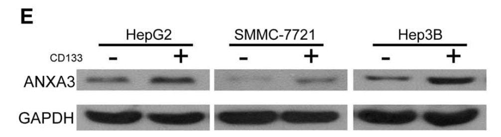
- Figure from citation: Western blot analysis of ANXA3 protein level by using anti-ANXA3 antibody in CD133+ HCC cell lines. HCC cells, including HepG2, SMMC-7721, and Hep3B cells. Dilution: 1:1000
- Validation comment
- WB
Supportive validation
- Submitted by
- OriGene (provider)
- Main image

- Experimental details
- Immunohistochemical staining of paraffin-embedded Human lung tissue within the normal limits using anti-ANXA3 mouse monoclonal antibody. (Heat-induced epitope retrieval by 10mM citric buffer, pH6.0, 100C for 10min, TA502058)
- Validation comment
- IHC

- Submitted by
- OriGene (provider)
- Main image

- Experimental details
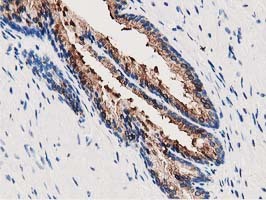
- Immunohistochemical staining of paraffin-embedded Human prostate tissue within the normal limits using anti-ANXA3 mouse monoclonal antibody. (Heat-induced epitope retrieval by 10mM citric buffer, pH6.0, 100C for 10min, TA502058)
- Validation comment
- IHC
- Submitted by
- OriGene (provider)
- Main image

- Experimental details
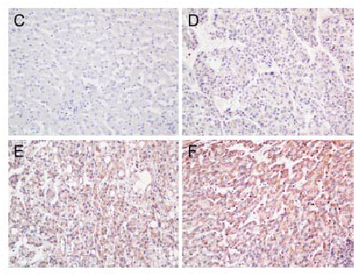
- Figure from citation: Representative images of immunohistochemical staining of ANXA3 expression in human liver tissues: (C) negative ANXA3 expression in non-tumorous liver parenchyma and (D) weak, (E) moderate, and (F) strong ANXA3 immunoreactivity in HCC tissues. Dilution: 1:500
- Validation comment
- IHC