Explore
 Validate
 Learn
Learn Western blot
Antibody data
- Antibody Data
- Antigen structure
- References [0]
- Comments [0]
- Validations
- Western blot [1]
- Immunocytochemistry [1]
- Immunohistochemistry [1]
- Flow cytometry [1]
Submit
Validation data
Reference
Comment
Report error
- Product number
- GTX81600 - Provider product page

- Provider
- GeneTex
- Proper citation
- GeneTex Cat#GTX81600, RRID:AB_11163340
- Product name
- ARGBP2 antibody, N-term
- Antibody type
- Polyclonal
- Reactivity
- Human
- Host
- Rabbit
No comments: Submit comment
Supportive validation
- Submitted by
- GeneTex (provider)
- Main image
- Experimental details
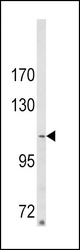
- Western blot analysis of ARGBP2 Antibody (N-term) (GTX81600) in MDA-MB231 cell line lysates (35ug/lane). ARGBP2 (arrow) was detected using the purified Pab.
- Validation comment
- WB
Supportive validation
- Submitted by
- GeneTex (provider)
- Main image
- Experimental details
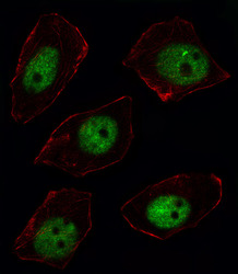
- Fluorescent image of U251 cell stained with ARGBP2 Antibody (N-term)(GTX81600).U251 cells were fixed with 4% PFA (20 min), permeabilized with Triton X-100 (0.1%, 10 min), then incubated with ARGBP2 primary antibody (1:25, 1 h at 37¢J). For secondary antibody, Alexa Fluor? 488 conjμgated donkey anti-rabbit antibody (green) was used (1:400, 50 min at 37¢J).Cytoplasmic actin was counterstained with Alexa Fluor? 555 (red) conjμgated Phalloidin (7units/ml, 1 h at 37¢J).ARGBP2 immunoreactivity is localized to Nucleus significantly.

Supportive validation
- Submitted by
- GeneTex (provider)
- Main image

- Experimental details
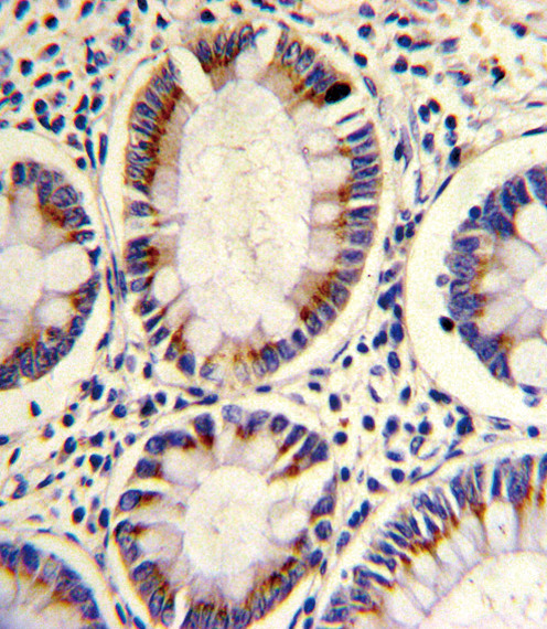
- Formalin-fixed and paraffin-embedded human colon carcinoma reacted with ARGBP2 Antibody (N-term)(GTX81600), which was peroxidase-conjμgated to the secondary antibody, followed by DAB staining. This data demonstrates the use of this antibody for immunohistochemistry; clinical relevance has not been evaluated.
Supportive validation
- Submitted by
- GeneTex (provider)
- Main image
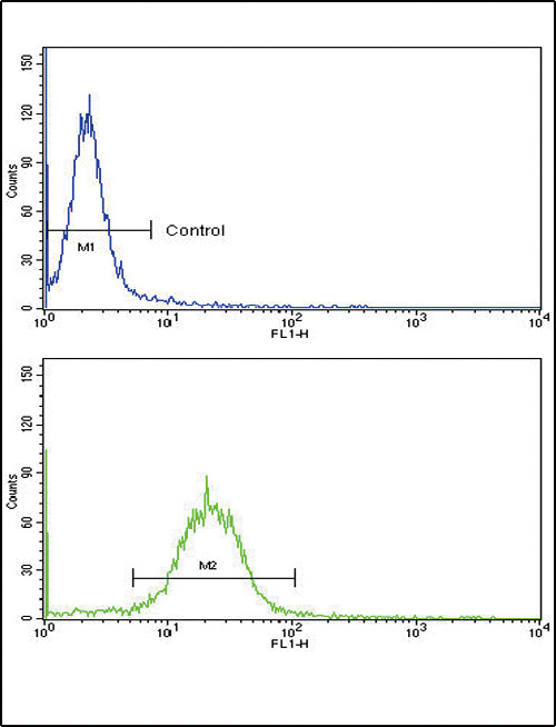
- Experimental details
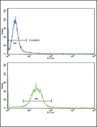
- Flow cytometric analysis of MDA-231 cells using ARGBP2 Antibody (N-term)(bottom histogram)(GTX81600) compared to a negative control cell (top histogram). FITC-conjμgated goat-anti-rabbit secondary antibodies were used for the analysis.