Explore
 Validate
 Learn
Learn Western blot
 ELISA
ELISAAntibody data
- Antibody Data
- Antigen structure
- References [3]
- Comments [0]
- Validations
- Western blot [2]
Submit
Validation data
Reference
Comment
Report error
- Product number
- NB100-1296 - Provider product page

- Provider
- Novus Biologicals
- Proper citation
- Novus Cat#NB100-1296, RRID:AB_2100905
- Product name
- Goat Polyclonal ETEA Antibody
- Antibody type
- Polyclonal
- Description
- Immunogen affinity purified.
- Reactivity
- Human, Mouse
- Host
- Goat
- Isotype
- IgG
- Vial size
- 0.1 mg
- Concentration
- 0.5 mg/ml
- Storage
- Store at -20C. Avoid freeze-thaw cycles.
Submitted references Sterol-induced dislocation of 3-hydroxy-3-methylglutaryl coenzyme A reductase from endoplasmic reticulum membranes into the cytosol through a subcellular compartment resembling lipid droplets.
Cloning and characterization of the highly expressed ETEA gene from blood cells of atopic dermatitis patients.
Cloning and characterization of the highly expressed ETEA gene from blood cells of atopic dermatitis patients.
Hartman IZ, Liu P, Zehmer JK, Luby-Phelps K, Jo Y, Anderson RG, DeBose-Boyd RA
The Journal of biological chemistry 2010 Jun 18;285(25):19288-98
The Journal of biological chemistry 2010 Jun 18;285(25):19288-98
Cloning and characterization of the highly expressed ETEA gene from blood cells of atopic dermatitis patients.
Imai Y, Nakada A, Hashida R, Sugita Y, Tanaka T, Tsujimoto G, Matsumoto K, Akasawa A, Saito H, Oshida T
Biochemical and biophysical research communications 2002 Oct 11;297(5):1282-90
Biochemical and biophysical research communications 2002 Oct 11;297(5):1282-90
No comments: Submit comment
Supportive validation
- Submitted by
- Novus Biologicals (provider)
- Main image
- Experimental details
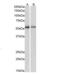
- Western Blot: ETEA Antibody [NB100-1296] - Staining of Human (A) and Mouse (B) Thymus lysate (35ug protein in RIPA buffer). Primary incubation was 1 hour. Detected by chemiluminescence.

- Submitted by
- Novus Biologicals (provider)
- Main image

- Experimental details
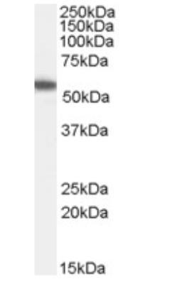
- Western Blot: ETEA Antibody [NB100-1296] - Staining (0.3ug/ml) of Jurkat lysate (RIPA buffer, 35ug total protein per lane). Primary incubated for 1 hour. Detected by chemiluminescence.