 Explore
 Validate
 Learn
Learn Western blot
Antibody data
- Antibody Data
- Antigen structure
- References [1]
- Comments [0]
- Validations
- Western blot [2]
- Immunocytochemistry [1]
- Immunohistochemistry [5]
Submit
Validation data
Reference
Comment
Report error
- Product number
- HPA017760 - Provider product page

- Provider
- Atlas Antibodies
- Proper citation
- Atlas Antibodies Cat#HPA017760, RRID:AB_2189993
- Product name
- Anti-SLFN5
- Antibody type
- Polyclonal
- Reactivity
- Human
- Host
- Rabbit
- Conjugate
- Unconjugated
- Antigen sequence
ERHGVGLDVPPIFRSHLDKMQKENHFLIFVKSWNT
EAGVPLATLCSNLYHRERTSTDVMDSQEALAFLKC
RTQTPTNINVSNSLGPQAAQGSVQYEGNINVSAAA
LFDRKRLQYLEKLNLPESTHVEFVMFSTDVSHCVK
D- Isotype
- IgG
- Vial size
- 100 µl
- Storage
- Store at +4°C for short term storage. Long time storage is recommended at -20°C.
Submitted references Role of Interferon (IFN )-inducible Schlafen-5 in Regulation of Anchorage-independent Growth and Invasion of Malignant Melanoma Cells
Katsoulidis E, Mavrommatis E, Woodard J, Shields M, Sassano A, Carayol N, Sawicki K, Munshi H, Platanias L
Journal of Biological Chemistry 2010 December;285(51):40333-40341
Journal of Biological Chemistry 2010 December;285(51):40333-40341
No comments: Submit comment
Supportive validation
Supportive validation
- Submitted by
- Atlas Antibodies (provider)
- Enhanced method
- Orthogonal validation
- Main image

- Experimental details
- Western blot analysis in human cell lines SK-MEL-30 and Caco-2 using Anti-SLFN5 antibody. Corresponding SLFN5 RNA-seq data are presented for the same cell lines. Loading control: Anti-HDAC1.

Supportive validation
- Submitted by
- Atlas Antibodies (provider)
- Main image
- Experimental details
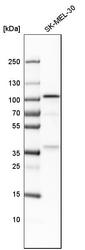
- Western blot analysis in human cell line SK-MEL-30.

Supportive validation
- Submitted by
- Atlas Antibodies (provider)
- Main image

- Experimental details
- Immunofluorescent staining of human cell line A-431 shows localization to nucleoplasm & vesicles.
- Sample type
- HUMAN

Supportive validation
- Submitted by
- Atlas Antibodies (provider)
- Main image

- Experimental details
- Immunohistochemical staining of human bone marrow shows strong nuclear positivity in subsets of hematopoietic cells.
- Sample type
- HUMAN

- Submitted by
- Atlas Antibodies (provider)
- Main image

- Experimental details
- Immunohistochemical staining of human Fallopian tube shows strong nuclear positivity in glandular cells.
- Sample type
- HUMAN

- Submitted by
- Atlas Antibodies (provider)
- Main image

- Experimental details
- Immunohistochemical staining of human lymph node shows strong nuclear positivity in non-germinal center cells.
- Sample type
- HUMAN

- Submitted by
- Atlas Antibodies (provider)
- Main image

- Experimental details
- Immunohistochemical staining of human endometrium shows strong nuclear positivity in glandular cells.
- Sample type
- HUMAN

- Submitted by
- Atlas Antibodies (provider)
- Main image

- Experimental details
- Immunohistochemical staining of human skeletal muscle shows weak nuclear positivity in myocytes as expected.
- Sample type
- HUMAN
