 Explore
 Validate
 Learn
Learn Western blot
 Immunohistochemistry
Antibody data
- Antibody Data
- Antigen structure
- References [3]
- Comments [0]
- Validations
- Western blot [1]
- Blocking/Neutralizing [1]
Submit
Validation data
Reference
Comment
Report error
- Product number
- AF653 - Provider product page

- Provider
- R&D Systems
- Product name
- Human CCL26/Eotaxin-3 Antibody
- Antibody type
- Polyclonal
- Description
- Antigen Affinity-purified. Detects CCL26/Eotaxin-3 in direct ELISAs and Western blots. In direct ELISAs, less than 5% cross-reactivity with recombinant human (rh) Eotaxin, recombinant mouse (rm) Eotaxin, rhEotaxin-2, and rmEotaxin-2 is observed.
- Reactivity
- Human
- Host
- Goat
- Conjugate
- Unconjugated
- Antigen sequence
Q9Y258- Isotype
- IgG
- Vial size
- 100 ug
- Concentration
- LYOPH
- Storage
- Use a manual defrost freezer and avoid repeated freeze-thaw cycles. 12 months from date of receipt, -20 to -70 °C as supplied. 1 month, 2 to 8 °C under sterile conditions after reconstitution. 6 months, -20 to -70 °C under sterile conditions after reconstitution.
Submitted references An initial investigation into endothelial CC chemokine expression in the human rheumatoid synovium.
Proton pump inhibitors decrease eotaxin-3 expression in the proximal esophagus of children with esophageal eosinophilia.
High levels of CCL26 in blister fluid and sera of patients with bullous pemphigoid.
Rump L, Mattey DL, Kehoe O, Middleton J
Cytokine 2017 Sep;97:133-140
Cytokine 2017 Sep;97:133-140
Proton pump inhibitors decrease eotaxin-3 expression in the proximal esophagus of children with esophageal eosinophilia.
Park JY, Zhang X, Nguyen N, Souza RF, Spechler SJ, Cheng E
PloS one 2014;9(7):e101391
PloS one 2014;9(7):e101391
High levels of CCL26 in blister fluid and sera of patients with bullous pemphigoid.
Kagami S, Kai H, Kakinuma T, Miyagaki T, Kamata M, Sugaya M, Tamaki K, Sato S
The Journal of investigative dermatology 2012 Jan;132(1):249-51
The Journal of investigative dermatology 2012 Jan;132(1):249-51
No comments: Submit comment
Supportive validation
- Submitted by
- R&D Systems (provider)
- Main image

- Experimental details
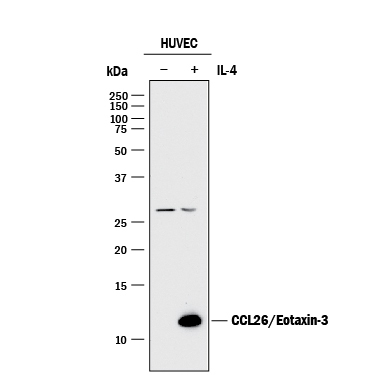
- Detection of Human CCL26/Eotaxin-3 by Western Blot. Western blot shows lysates of HUVEC human umbilical vein endothelial cells untreated (-) or treated (+) with 100 U/mL Recombinant Human IL-4 (Catalog # 204-IL) for 24 hours. PVDF membrane was probed with 1 µg/mL of Goat Anti-Human CCL26/Eotaxin-3 Antigen Affinity-purified Polyclonal Antibody (Catalog # AF653) followed by HRP-conjugated Anti-Goat IgG Secondary Antibody (Catalog # HAF017). A specific band was detected for CCL26/ Eotaxin-3 at approximately 12 kDa (as indicated). This experiment was conducted under reducing conditions and using Immunoblot Buffer Group 1.
Supportive validation
- Submitted by
- R&D Systems (provider)
- Main image
- Experimental details
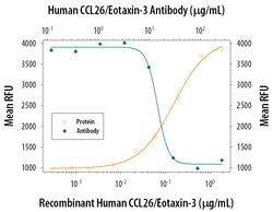
- Chemotaxis Induced by CCL26/Eotaxin-3 and Neutralizatio by Human CCL26/Eotaxin-3 Antibody. Recombinant Human CCL26/Eotaxin-3 (Catalog # 346-E3) chemoattracts the BaF3 mouse pro-B cell line transfected with mouse CCR3 in a dose-dependent manner (orange line). The amount of cells that migrated through to the lower chemotaxis chamber was measured by Resazurin (Catalog # AR002). Chemotaxis elicited by Recombinant Human CCL26/Eotaxin-3 (1 µg/mL) is neutralized (green line) by increasing concentrations of Human CCL26/Eotaxin-3 Antigen Affinity-purified Polyclonal Antibody (Catalog # AF653). The ND50 is typically 10-25 µg/mL.
