Explore
 Validate
 Learn
Learn Immunocytochemistry
Antibody data
- Antibody Data
- Antigen structure
- References [0]
- Comments [0]
- Validations
- Immunocytochemistry [6]
- Chromatin Immunoprecipitation [1]
Submit
Validation data
Reference
Comment
Report error
- Product number
- MA5-44597 - Provider product page

- Provider
- Invitrogen Antibodies
- Product name
- NSD1 Recombinant Rabbit Monoclonal Antibody (1B1)
- Antibody type
- Monoclonal
- Antigen
- Recombinant full-length protein
- Description
- Predicted band size: 297 kDa. Positive Control: HEK293. Subcellular Location: Nucleus.
- Reactivity
- Human
- Host
- Rabbit
- Isotype
- IgG
- Antibody clone number
- 1B1
- Vial size
- 100 µL
- Concentration
- 1 mg/mL
- Storage
- Store at 4°C short term. For long term storage, store at -20°C, avoiding freeze/thaw cycles.
No comments: Submit comment
Supportive validation
- Submitted by
- Invitrogen Antibodies (provider)
- Main image
- Experimental details
- Immunofluorescence analysis of NSD1 using 293 cells. The cells were fixed 4% paraformaldehyde for 10 minutes, permeabilized with 0.05% Triton™ X-100 in PBS for 20 minutes, and blocked with 2% negative goat serum for 30 minutes at room temperature. The cells were labeled with NSD1 Recombinant Rabbit Monoclonal Antibody (1B1) (Product # MA5-44597) at 1:100 dilution in 0.1% BSA for 1 hour at room temperature and then with Alexa Fluor®488 Goat anti-Rabbit IgG secondary antibody (1:1,000 dilution) for 1 hour at room temperature (green). Nuclei were stained with DAPI (blue). The images were captured at 200X magnification.
- Submitted by
- Invitrogen Antibodies (provider)
- Main image
- Experimental details
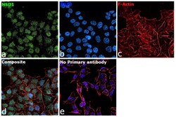
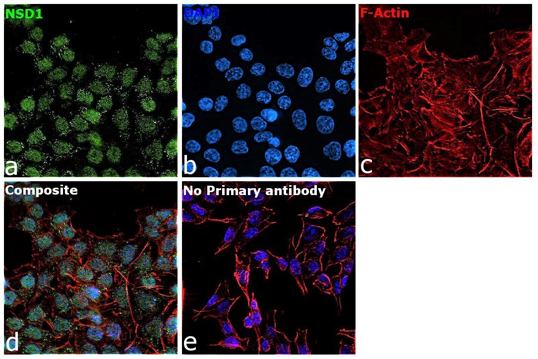
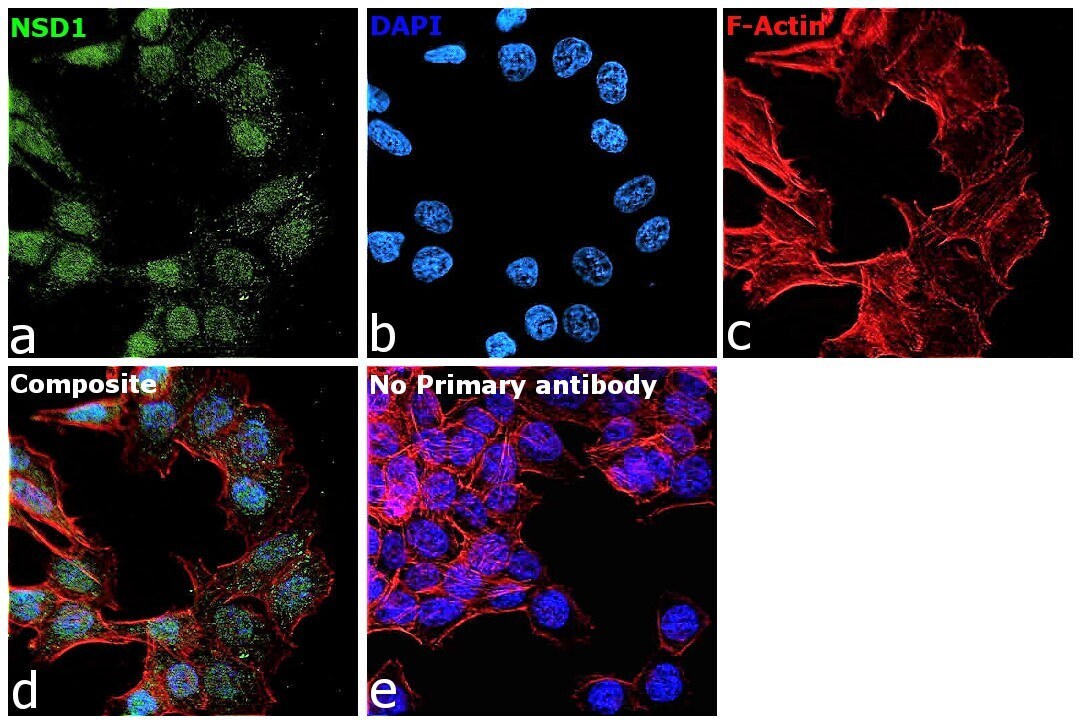
- Immunofluorescence analysis of NSD1 was performed using 70% confluent log phase U-2 OS cells. The cells were fixed with 4% paraformaldehyde for 10 minutes, permeabilized with 0.1% Triton™ X-100 for 15 minutes, and blocked with 2% BSA for 1 hour at room temperature. The cells were labeled with NSD1 Recombinant Rabbit Monoclonal Antibody (1B1) (Product # MA5-44597) at 1:100 dilution in 0.1% BSA, incubated at 4 degree celsius overnight and then labeled with Donkey anti-Rabbit IgG (H+L) Highly Cross-Adsorbed Secondary Antibody, Alexa Fluor Plus 488 (Product # A32790), (1:2,000 dilution), for 45 minutes at room temperature (Panel a: Green). Nuclei (Panel b:Blue) were stained with ProLong™ Diamond Antifade Mountant with DAPI (Product # P36962). F-actin (Panel c: Red) was stained with Rhodamine Phalloidin (Product # R415, 1:300 dilution). Panel d represents the merged image showing nuclear localization. Panel e represents control cells with no primary antibody to assess background. The images were captured at 60X magnification.
- Submitted by
- Invitrogen Antibodies (provider)
- Main image
- Experimental details
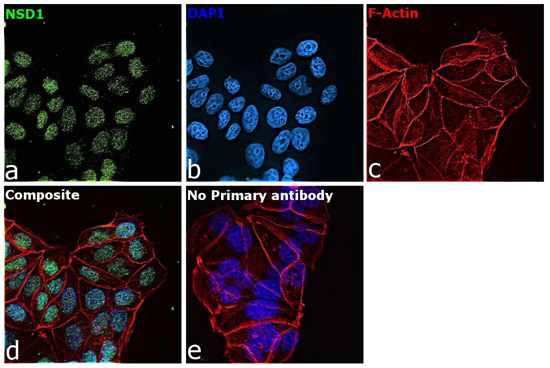
- Immunofluorescence analysis of NSD1 was performed using 70% confluent log phase MCF-7 cells. The cells were fixed with 4% paraformaldehyde for 10 minutes, permeabilized with 0.1% Triton™ X-100 for 15 minutes, and blocked with 2% BSA for 1 hour at room temperature. The cells were labeled with NSD1 Recombinant Rabbit Monoclonal Antibody (1B1) (Product # MA5-44597) at 1:100 dilution in 0.1% BSA, incubated at 4 degree celsius overnight and then labeled with Donkey anti-Rabbit IgG (H+L) Highly Cross-Adsorbed Secondary Antibody, Alexa Fluor Plus 488 (Product # A32790), (1:2,000 dilution), for 45 minutes at room temperature (Panel a: Green). Nuclei (Panel b:Blue) were stained with ProLong™ Diamond Antifade Mountant with DAPI (Product # P36962). F-actin (Panel c: Red) was stained with Rhodamine Phalloidin (Product # R415, 1:300 dilution). Panel d represents the merged image showing nuclear localization. Panel e represents control cells with no primary antibody to assess background. The images were captured at 60X magnification.
- Submitted by
- Invitrogen Antibodies (provider)
- Main image
- Experimental details
- Immunofluorescence analysis of NSD1 was performed using 70% confluent log phase HCT 116 cells. The cells were fixed with 4% paraformaldehyde for 10 minutes, permeabilized with 0.1% Triton™ X-100 for 15 minutes, and blocked with 2% BSA for 1 hour at room temperature. The cells were labeled with NSD1 Recombinant Rabbit Monoclonal Antibody (1B1) (Product # MA5-44597) at 1:100 dilution in 0.1% BSA, incubated at 4 degree celsius overnight and then labeled with Donkey anti-Rabbit IgG (H+L) Highly Cross-Adsorbed Secondary Antibody, Alexa Fluor Plus 488 (Product # A32790), (1:2,000 dilution), for 45 minutes at room temperature (Panel a: Green). Nuclei (Panel b:Blue) were stained with ProLong™ Diamond Antifade Mountant with DAPI (Product # P36962). F-actin (Panel c: Red) was stained with Rhodamine Phalloidin (Product # R415, 1:300 dilution). Panel d represents the merged image showing nuclear localization. Panel e represents control cells with no primary antibody to assess background. The images were captured at 60X magnification.
- Submitted by
- Invitrogen Antibodies (provider)
- Main image
- Experimental details
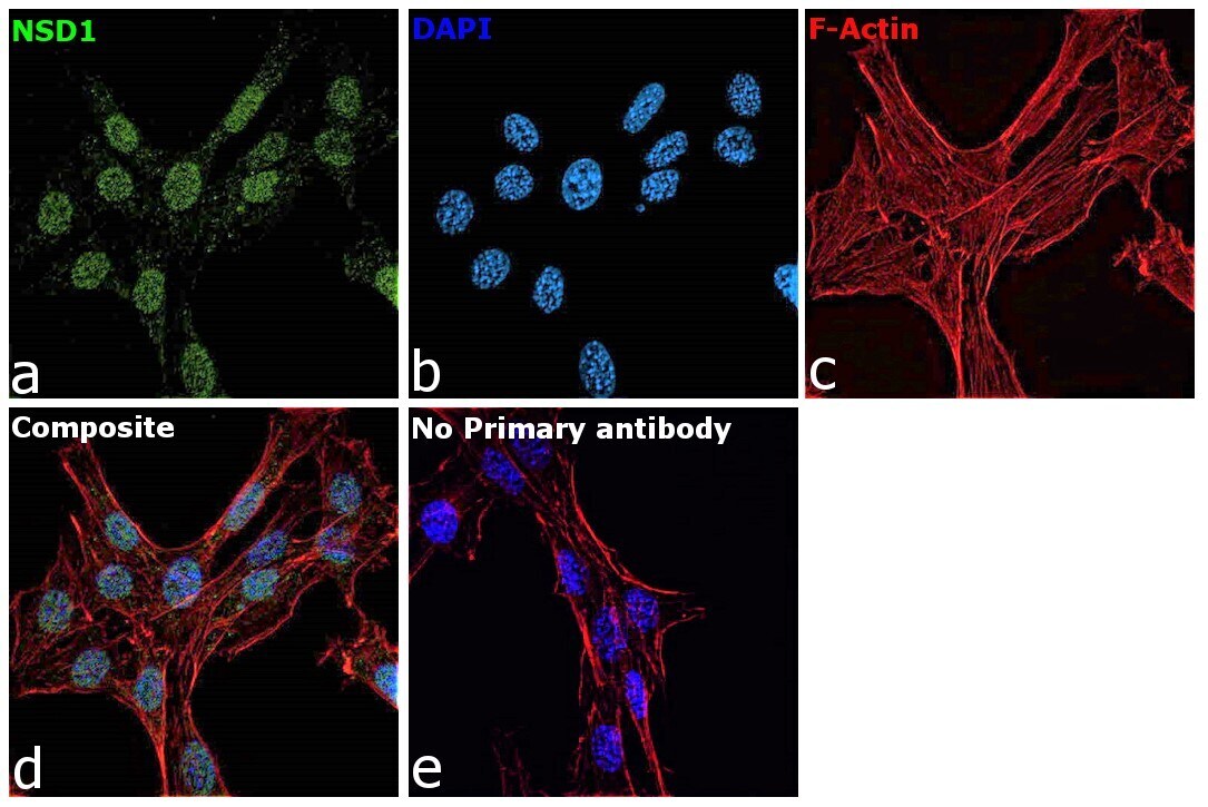
- Immunofluorescence analysis of NSD1 was performed using 70% confluent log phase NIH 3T3 cells. The cells were fixed with 4% paraformaldehyde for 10 minutes, permeabilized with 0.1% Triton™ X-100 for 15 minutes, and blocked with 2% BSA for 1 hour at room temperature. The cells were labeled with NSD1 Recombinant Rabbit Monoclonal Antibody (1B1) (Product # MA5-44597) at 1:100 dilution in 0.1% BSA, incubated at 4 degree celsius overnight and then labeled with Donkey anti-Rabbit IgG (H+L) Highly Cross-Adsorbed Secondary Antibody, Alexa Fluor Plus 488 (Product # A32790), (1:2,000 dilution), for 45 minutes at room temperature (Panel a: Green). Nuclei (Panel b:Blue) were stained with ProLong™ Diamond Antifade Mountant with DAPI (Product # P36962). F-actin (Panel c: Red) was stained with Rhodamine Phalloidin (Product # R415, 1:300 dilution). Panel d represents the merged image showing nuclear localization. Panel e represents control cells with no primary antibody to assess background. The images were captured at 60X magnification.
- Submitted by
- Invitrogen Antibodies (provider)
- Main image
- Experimental details
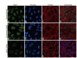
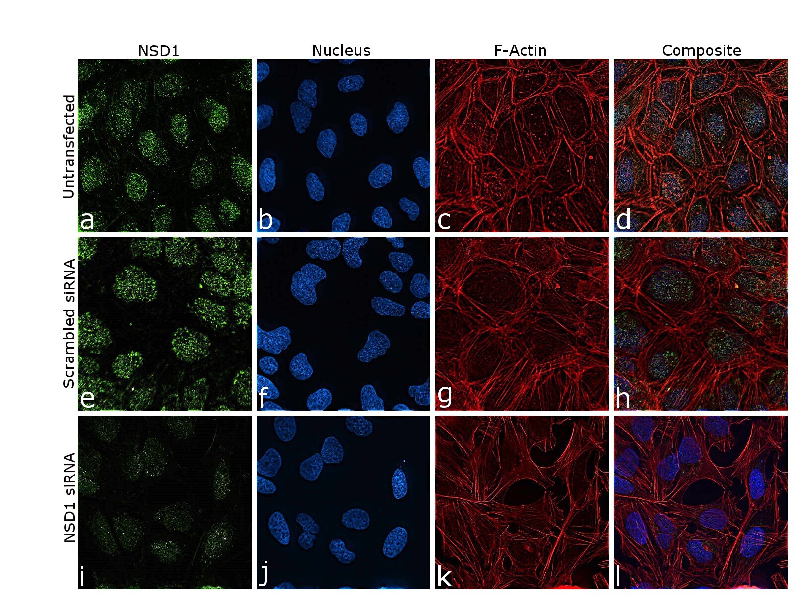
- Knockdown of NSD1 was achieved by transfecting U-2 OS cells with NSD1 specific siRNA (Silencer® select Product # s34630, s34631). Immunofluorescence analysis was performed on U-2 OS cells (untransfected, panels a-d), transfected with non-specific scrambled siRNA (panels e-h) and transfected with NSD1 specific siRNA (panels i-l). Cells were fixed, permeabilized, and labelled with NSD1 Recombinant Rabbit Monoclonal Antibody (1B1) (Product # MA5-44597, 1:100 dilution), followed by Donkey anti-Rabbit IgG (H+L) Highly Cross-Adsorbed Secondary Antibody, Alexa Fluor Plus 488, Product # A32790), 1:2,000 dilution). Nuclei (blue) were stained using ProLong™ Diamond Antifade Mountant with DAPI (Product # P36962), and Rhodamine Phalloidin (Product # R415, 1:300 dilution) was used for cytoskeletal F-actin (red) staining. Reduction of specific signal was observed upon siRNA mediated knockdown (panels i-l) confirming specificity of the antibody to NSD1 (green). The images were captured at 60X magnification.
Supportive validation
- Submitted by
- Invitrogen Antibodies (provider)
- Main image
- Experimental details
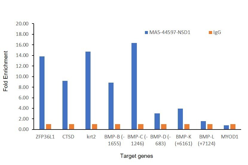
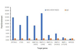
- Chromatin Immunoprecipitation (ChIP) was performed using NSD1 Recombinant Rabbit Monoclonal Antibody (1B1) (Product # MA5-44597) at 5 µg on sheared chromatin from U-2 OS cells using the MAGnify ChIP System (Product #49-2024). Normal Rabbit IgG was used as a negative IP control. The purified DNA was analyzed by qPCR with PCR primer pairs over ZFP36L1 (active), CTSD (active), KRT2 (active) and MYOD1 (inactive). We further analyzed the binding of NSD1 to different loci of BMP4 gene- Loci B region (-1655 bp) (active), C region (-1246 bp) (active), D region (-683 bp) (inactive), K region (+6161 bp) (inactive), L region (+7124 bp) (inactive) for NSD1. Antibody specificity was demonstrated by detection of enrichment of the target protein at specific gene loci. Data is presented as fold enrichment of the antibody signal versus the Rabbit Isotype using the comparative CT method.