Explore
 Validate
 Learn
Learn Western blot
Antibody data
- Antibody Data
- Antigen structure
- References [0]
- Comments [0]
- Validations
- Western blot [1]
- Immunocytochemistry [1]
- Immunohistochemistry [1]
Submit
Validation data
Reference
Comment
Report error
- Product number
- GTX16973 - Provider product page

- Provider
- GeneTex
- Product name
- SCN1B antibody
- Antibody type
- Polyclonal
- Reactivity
- Mouse, Rat
- Host
- Rabbit
No comments: Submit comment
Supportive validation
- Submitted by
- GeneTex (provider)
- Main image

- Experimental details
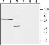
- Anti-NaVbeta1_(extracellular) - Western blot analysis of rat brain (lanes 1 and 4), mouse brain (lanes 2 and 5) and rat skeletal muscle (lanes 3 and 6): 1-3. Anti-NaV£]1 (extracellular) antibody, (1:400). 4-6. Anti-NaV£]1 (extracellular) antibody, preincubated with the control peptide antigen.
Supportive validation
- Submitted by
- GeneTex (provider)
- Main image
- Experimental details
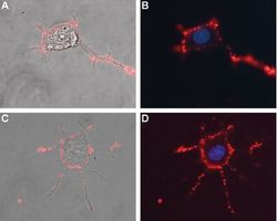
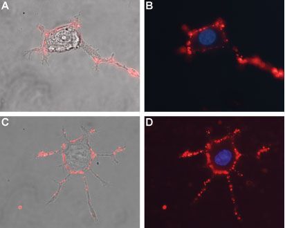
- Anti-NaVbeta1_(extracellular) - Expression of NaVbeta1 in differentiated rat PC12 cells Immunocytochemical staining of live and intact differentiated rat PC12 cells. PC12 differentiation was induced by mNGF 2.5S (>95%). A, C. Merge of NaV£]1 staining using Anti-NaV£]1 (extracellular) antibody, (1:50), (red) with live cell imaging. B, D. Merge of NaV£]1 staining (1:50), (red) with nuclear staining using DAPI as the counterstain.
Supportive validation
- Submitted by
- GeneTex (provider)
- Main image

- Experimental details
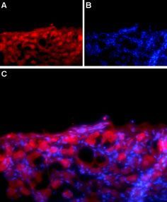
- Anti-NaVbeta1_(extracellular) - Expression of NaVbeta1 in rat DRG Immunohistochemical staining of adult rat dorsal root ganglion (DRG) using Anti- NaV£]1 (extracellular) antibody followed by goat anti-rabbit-AlexaFluor-594 secondary antibody. A. Nav£]1 labeling (red) appears in the cell bodies of the DRG neurons. B. Nuclear staining using DAPI as the counterstain (blue). C. Merged image of A and B.