Explore
 Validate
 Learn
Learn Western blot
 Immunohistochemistry
Antibody data
- Antibody Data
- Antigen structure
- References [2]
- Comments [0]
- Validations
- Immunohistochemistry [2]
Submit
Validation data
Reference
Comment
Report error
- Product number
- NB100-1620 - Provider product page

- Provider
- Novus Biologicals
- Proper citation
- Novus Cat#NB100-1620, RRID:AB_10003258
- Product name
- Rabbit Polyclonal mu Opioid R/OPRM1 Antibody
- Antibody type
- Polyclonal
- Description
- Unpurified. Mu Opioid Receptor
- Reactivity
- Human, Mouse, Rat, Simian
- Host
- Rabbit
- Isotype
- IgG
- Vial size
- 0.05 ml
- Storage
- Store at -20C. Avoid freeze-thaw cycles.
Submitted references Rules to activate CD8+T cells through regulating subunits of opioid receptors by methionine enkephalin (MENK).
Exposure to opiates in female adolescents alters mu opiate receptor expression and increases the rewarding effects of morphine in future offspring.
Jiao X, Wang X, Wang R, Geng J, Liu N, Chen H, Griffin N, Shan F
International immunopharmacology 2018 Dec;65:76-83
International immunopharmacology 2018 Dec;65:76-83
Exposure to opiates in female adolescents alters mu opiate receptor expression and increases the rewarding effects of morphine in future offspring.
Vassoler FM, Wright SJ, Byrnes EM
Neuropharmacology 2016 Apr;103:112-21
Neuropharmacology 2016 Apr;103:112-21
No comments: Submit comment
Supportive validation
- Submitted by
- Novus Biologicals (provider)
- Main image

- Experimental details
- Immunohistochemistry: mu Opioid R/OPMR1 Antibody [NB100-1620] - Striatum

- Submitted by
- Novus Biologicals (provider)
- Main image

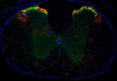
- Experimental details
- Immunohistochemistry-Frozen: mu Opioid R/OPRM1 Antibody [NB100-1620] - Opioid R/OPRM1 (green) and Substance P (red) were detected in frozen mouse spinal cord and DRG tissue by fluorescent multiplex IHC. Mu Opioid R/OPRM1 Antibody (NB100-1620), used at 1:100, and Substance P Antibody (NB100-65219), used at 1:50, were applied to the tissue and simultaneously incubated at 4C overnight. Anti-Rabbit Alexa Fluor(R) 488-conjugated secondary antibody and anti-Rat NL557-conjugated secondary antibody (NL013) were applied to the tissue, and simultaneously incubated at room temperature, protected from light for 1 hour. Coverslips were applied to the tissue slide along with mounting media containing DAPI (blue) and imaged using a 4X objective lens.