 Explore
 Validate
 Learn
Learn Western blot
 ELISA
ELISAAntibody data
- Antibody Data
- Antigen structure
- References [3]
- Comments [0]
- Validations
- Western blot [2]
- Immunohistochemistry [1]
Submit
Validation data
Reference
Comment
Report error
- Product number
- ABIN359509 - Provider product page

- Provider
- antibodies-online
- Product name
- anti-NEDD8 Activating Enzyme E1 Subunit 1 (NAE1) (C-Term) antibody
- Antibody type
- Polyclonal
- Antigen
- This antibody is generated from rabbits immunized with a KLH conjugated synthetic peptide selected from the C-terminal region of human APPBP1.
- Description
- Protein G column, eluted with high and low pH buffers and neutralized immediately, followed by dialysis against PBS.
- Reactivity
- Human
- Host
- Rabbit
- Epitope
- C-Term
- Vial size
- 0.4 mL
- Concentration
- 0.25 mg/mL
- Storage
- Store the antibody at 2 - 8°C up to one month or (in aliquots) at -20°C for longer.
- Handling
- Avoid repeated freezing and thawing.
Submitted references APP-BP1 mediates APP-induced apoptosis and DNA synthesis and is increased in Alzheimer's disease brain.
ASPP2 inhibits APP-BP1-mediated NEDD8 conjugation to cullin-1 and decreases APP-BP1-induced cell proliferation and neuronal apoptosis.
Insights into the ubiquitin transfer cascade from the structure of the activating enzyme for NEDD8.
Chen Y, Liu W, McPhie DL, Hassinger L, Neve RL
The Journal of cell biology 2003 Oct 13;163(1):27-33
The Journal of cell biology 2003 Oct 13;163(1):27-33
ASPP2 inhibits APP-BP1-mediated NEDD8 conjugation to cullin-1 and decreases APP-BP1-induced cell proliferation and neuronal apoptosis.
Chen Y, Liu W, Naumovski L, Neve RL
Journal of neurochemistry 2003 May;85(3):801-9
Journal of neurochemistry 2003 May;85(3):801-9
Insights into the ubiquitin transfer cascade from the structure of the activating enzyme for NEDD8.
Walden H, Podgorski MS, Schulman BA
Nature 2003 Mar 20;422(6929):330-4
Nature 2003 Mar 20;422(6929):330-4
No comments: Submit comment
Supportive validation
- Submitted by
- antibodies-online (provider)
- Main image
- Experimental details
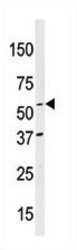
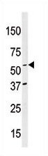
- Western blot analysis of anti-APPBP1 (C-term) (AP13993PU-N) in mouse brain tissue lysates (35 μg/lane). APPBP1(arrow) was detected using the purified Pab (1:60 dilution).
- Submitted by
- antibodies-online (provider)
- Main image
- Experimental details
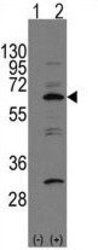
- Western blot analysis of APP-BP1(arrow) using rabbit polyclonal APP-BP1 Antibody (C-term) (AP13993PU-N). 293 cell lysates (2 μg/lane) either nontransfected (Lane 1) or transiently transfected with the APP-BP1 gene (Lane 2) (Origene Technologies).

Supportive validation
- Submitted by
- antibodies-online (provider)
- Main image

- Experimental details
- Formalin-fixed and paraffin-embedded human breast carcinoma reacted with anti-APPBP1 (C-term), which was peroxidase-conjugated to the secondary antibody, followed by DAB staining. This data demonstrates the use of this antibody for immunohistochemistry; clinical relevance has not been evaluated.
