 Explore
 Validate
 Learn
LearnPA5-47869
antibody from Invitrogen Antibodies
Targeting: CELSR2
ADGRC2, CDHF10, EGFL2, Flamingo1, KIAA0279, MEGF3
 Western blot
Antibody data
- Antibody Data
- Antigen structure
- References [0]
- Comments [0]
- Validations
- Western blot [2]
- Immunohistochemistry [1]
- Flow cytometry [4]
Submit
Validation data
Reference
Comment
Report error
- Product number
- PA5-47869 - Provider product page

- Provider
- Invitrogen Antibodies
- Product name
- CELSR2 Polyclonal Antibody
- Antibody type
- Polyclonal
- Antigen
- Recombinant full-length protein
- Reactivity
- Human, Mouse
- Host
- Goat
- Isotype
- IgG
- Vial size
- 100 µg
- Concentration
- 0.2 mg/mL
- Storage
- -20° C, Avoid Freeze/Thaw Cycles
No comments: Submit comment
Supportive validation
- Submitted by
- Invitrogen Antibodies (provider)
- Main image
- Experimental details
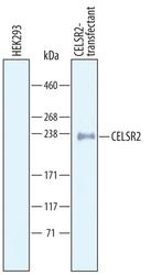
- Western blot analysis from lysates of HEK293 human embryonic kidney cell line either mock transfected or transfected with human CELSR2. PVDF Membrane was probed with 1 µg/mL of Goat Anti-human CELSR2 Antigen Affinity-purified Polyclonal Antibody (Product # PA5-47869) followed by HRP-conjugated Anti-Goat IgG Secondary Antibody. A specific band was detected for CELSR2 at approximately 240 kDa (as indicated). This experiment was conducted under reducing conditions.

- Submitted by
- Invitrogen Antibodies (provider)
- Main image

- Experimental details
- Western blot analysis of CELSR2 in HEK293 human embryonic kidney cell line either mock transfected or transfected with human CELSR2. Samples were incubated in CELSR2 polyclonal antibody (Product # PA5-47869) using a dilution of 1 µg/mL followed by a HRP-conjugated Anti-Goat IgG secondary antibody. A specific band was detected for CELSR2 at approximately 240 kDa (as indicated). This experiment was conducted under reducing conditions.

Supportive validation
- Submitted by
- Invitrogen Antibodies (provider)
- Main image
- Experimental details
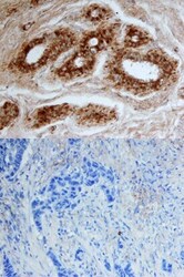
- Immunohistochemical analysis of CELSR2 in immersion fixed paraffin-embedded sections of human breast. Samples were incubated in CELSR2 polyclonal antibody (Product # PA5-47869) using a dilution of 10 µg/mL overnight at 4 °C. Tissue was stained using the Anti-Goat HRP-DAB Cell & Tissue Staining Kit (brown) and counterstained with hematoxylin (blue). Lower panel shows a lack of labeling when primary antibodies are omitted and tissue is stained only with secondary antibody followed by incubation with detection reagents. Specific staining was localized to ductal epithelium.

Supportive validation
- Submitted by
- Invitrogen Antibodies (provider)
- Main image

- Experimental details
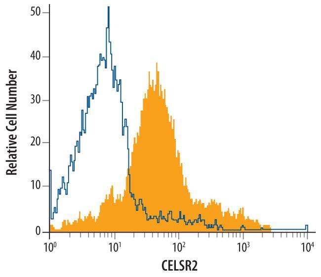
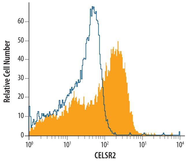
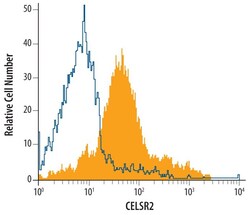
- Flow cytometric analysis of SH-SY5Y human neuroblastoma cell line was stained with Goat Anti-human CELSR2 Antigen Affinity-purified Polyclonal Antibody (Product # PA5-47869, filled histogram) or isotype control antibodyopen histogram), followed by Allophycocyanin-conjugated Anti-Goat IgG Secondary Antibody.
- Submitted by
- Invitrogen Antibodies (provider)
- Main image

- Experimental details
- Flow cytometric analysis of SH-SY5Y human neuroblastoma cell line was stained with Goat Anti-human CELSR2 Antigen Affinity-purified Polyclonal Antibody (Product # PA5-47869, filled histogram) or isotype control antibodyopen histogram), followed by Allophycocyanin-conjugated Anti-Goat IgG Secondary Antibody.
- Submitted by
- Invitrogen Antibodies (provider)
- Main image
- Experimental details
- Flow cytometry of CELSR2 in SH‚SY5Y human neuroblastoma cell line. Samples were incubated in CELSR2 polyclonal antibody (Product # PA5-47869) or isotype control antibody followed by Allophycocyanin-conjugated Anti-Goat IgG Secondary Antibody.

- Submitted by
- Invitrogen Antibodies (provider)
- Main image

- Experimental details
- Flow cytometry of CELSR2 in bEnd.3 mouse endothelioma cell line. Samples were incubated in CELSR2 polyclonal antibody (Product # PA5-47869) or isotype control antibody followed by Allophycocyanin-conjugated Anti-Goat IgG Secondary Antibody.
