Explore
 Validate
 Learn
Learn Western blot
Antibody data
- Antibody Data
- Antigen structure
- References [0]
- Comments [0]
- Validations
- Western blot [1]
- Immunohistochemistry [2]
- Flow cytometry [1]
Submit
Validation data
Reference
Comment
Report error
- Product number
- NBP2-01853 - Provider product page

- Provider
- Novus Biologicals
- Product name
- Mouse Monoclonal RAMP2 Antibody
- Antibody type
- Monoclonal
- Description
- Affinity purified.
- Reactivity
- Human, Simian
- Host
- Mouse
- Isotype
- IgG
- Vial size
- 0.1 ml
- Concentration
- 1.25 mg/ml
- Storage
- Store at -20C. Avoid freeze-thaw cycles.
No comments: Submit comment
Supportive validation
- Submitted by
- Novus Biologicals (provider)
- Main image
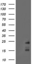
- Experimental details
- Western Blot: RAMP2 Antibody (4E5) [NBP2-01853] - HEK293T cells were transfected with the pCMV6-ENTRY control (Left lane) or pCMV6-ENTRY RAMP2 (Right lane) cDNA for 48 hrs and lysed. Equivalent amounts of cell lysates (5 ug per lane) were separated by SDS-PAGE and immunoblotted with anti-RAMP2.

Supportive validation
- Submitted by
- Novus Biologicals (provider)
- Main image

- Experimental details
- Immunohistochemistry-Paraffin: RAMP2 Antibody (4E5) [NBP2-01853] - Staining of paraffin-embedded Human Kidney tissue using anti-RAMP2 mouse monoclonal antibody.

- Submitted by
- Novus Biologicals (provider)
- Main image

- Experimental details
- Immunohistochemistry-Paraffin: RAMP2 Antibody (4E5) [NBP2-01853] - FFPE gingival tissue from Macaca mulatta was stained with RAMP2 Antibody (4E5) as described in "Other experimental details". Clear membranous and cytoplasmic staining is seen in the epithelium and also in the inflammatory cells infiltrating the stroma. Antibody diluted 1:150. This image was submitted via customer Review.

Supportive validation
- Submitted by
- Novus Biologicals (provider)
- Main image

- Experimental details
- Flow Cytometry: RAMP2 Antibody (4E5) [NBP2-01853] - HEK293T cells transfected with either overexpression plasmid (Red) or empty vector control plasmid (Blue) were immunostained by anti-RAMP2 antibody, and then analyzed by flow cytometry.