 Explore
 Validate
 Learn
Learn Western blot
Antibody data
- Antibody Data
- Antigen structure
- References [0]
- Comments [0]
- Validations
- Western blot [2]
- Immunocytochemistry [1]
- Flow cytometry [2]
Submit
Validation data
Reference
Comment
Report error
- Product number
- AP17302PU-N - Provider product page

- Provider
- Acris Antibodies GmbH
- Proper citation
- Acris Antibodies GmbH Cat#AP17302PU-N, RRID:AB_10652864
- Product name
- anti Endothelin A receptor
- Antibody type
- Polyclonal
- Antigen
- KLH conjugated synthetic peptide selected between aa 132-162 amino acids from the Center region of Human EDNRA
- Reactivity
- Human
- Host
- Rabbit
- Vial size
- 0.4 ml
- Concentration
- 2 mg/mL
No comments: Submit comment
Supportive validation
- Submitted by
- Acris Antibodies GmbH (provider)
- Main image
- Experimental details
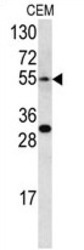
- Western blot analysis of EDNRA antibody (Center) (AP17302PU-N) in CEM cell line lysates (35 µg/lane). EDNRA (arrow) was detected using the purified Pab.

- Submitted by
- Acris Antibodies GmbH (provider)
- Main image

- Experimental details
- Western blot Analysis of EDNRA Antibody (Center) Cat.-No AP17302PU-NÂ in CEM cell line lysates (35ug/lane). EDNRA (arrow) was detected using the purified Pab.

Supportive validation
- Submitted by
- Acris Antibodies GmbH (provider)
- Main image
- Experimental details
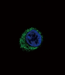
- Confocal Immunofluorescent Analysis of EDNRA Antibody (Center) Cat.-No AP17302PU-N with HepG2 cell followed by Alexa Fluor488-conjugated Goat anti-Rabbit lgG (green). DAPI was used to stain the cell nuclear (blue).

Supportive validation
- Submitted by
- Acris Antibodies GmbH (provider)
- Main image

- Experimental details
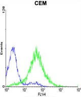
- Flow cytometric analysis of CEM cells using EDNRA Antibody (Center) (green histogram) compared to a negative control cell (blue histogram). FITC-conjugated goat-anti-rabbit secondary antibodies were used for the analysis.
- Submitted by
- Acris Antibodies GmbH (provider)
- Main image
- Experimental details
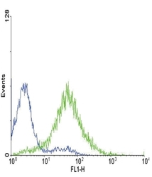
- Flow Cytometric Analysis of CEM cells using EDNRA Antibody (Center) Cat.-No AP17302PU-NÂ (green histogram) compared to a negative control cell (blue histogram). FITC-conjugated Goat-anti-Rabbit secondary antibodies were used for the analysis.
