Explore
 Validate
 Learn
Learn Western blot
Antibody data
- Antibody Data
- Antigen structure
- References [1]
- Comments [0]
- Validations
- Western blot [2]
- Immunohistochemistry [3]
- Flow cytometry [1]
- Other assay [1]
Submit
Validation data
Reference
Comment
Report error
- Product number
- PA5-95277 - Provider product page

- Provider
- Invitrogen Antibodies
- Product name
- CYP1B1 Polyclonal Antibody
- Antibody type
- Polyclonal
- Antigen
- Recombinant full-length protein
- Reactivity
- Human, Mouse, Rat
- Host
- Rabbit
- Isotype
- IgG
- Vial size
- 100 µg
- Concentration
- 500 µg/mL
- Storage
- Store at 4°C short term. For long term storage, store at -20°C, avoiding freeze/thaw cycles.
Submitted references Attenuation of Polycyclic Aromatic Hydrocarbon (PAH)-Mediated Pulmonary DNA Adducts and Cytochrome P450 (CYP)1B1 by Dietary Antioxidants, Omega-3 Fatty Acids, in Mice.
Zhou G, Jiang W, Xia G, Wang L, Richardson M, Chu C, Moorthy B
Antioxidants (Basel, Switzerland) 2022 Jan 5;11(1)
Antioxidants (Basel, Switzerland) 2022 Jan 5;11(1)
No comments: Submit comment
Supportive validation
- Submitted by
- Invitrogen Antibodies (provider)
- Main image
- Experimental details
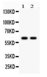
- Western blot analysis of CYP1B1 in Lane 1: rat kidney tissue lysate Lane 2: MCF-7 whole cell lysate After Electrophoresis, proteins were transferred to a Nitrocellulose membrane at 150mA for 50-90 minutes. Electrophoresis was performed with 5-20% SDS-PAGE gel (70V, Stacking gel; 90V Resolving gel, Time: 2-3 hours), transferred to a nitrocellulose membrane and blocked using 5% Non-fat Milk/TBS (1.5 hrs at room temperature). Samples were incubated with CYP1B1 polyclonal antibody (Product # PA5-95277) using a 0.5 µg/mL dilution, followed by a goat anti-rabbit IgG-HRP at a dilution of 1:10,000, and developed with enhanced chemiluminescence (ECL).

- Submitted by
- Invitrogen Antibodies (provider)
- Main image
- Experimental details
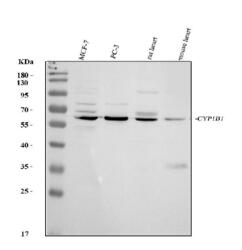
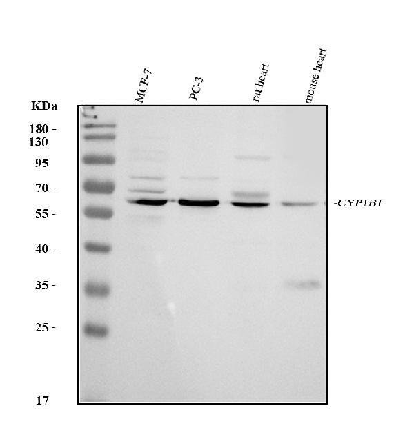
- Western blot analysis of CYP1B1 in, Lane 1: human MCF-7 whole cell lysates, Lane 2: human PC-3 whole cell lysates, Lane 3: rat heart tissue lysates, Lane 4: mouse heart tissue lysates. Electrophoresis was performed on a 5-20% SDS-PAGE gel at 70V (Stacking gel) / 90V (Resolving gel) for 2-3 hours. The sample well of each lane was loaded with 30 µg of sample under reducing conditions. After Electrophoresis, proteins were transferred to a nitrocellulose membrane at 150 mA for 50-90 minutes. The membrane was blocked with 5% non-fat milk/TBS for 1. 5 hour at RT. The membrane was incubated with CYP1B1 Polyclonal Antibody (Product # PA5-95277) at 0.5 μg/mL overnight at 4°C, then washed with TBS-0. 1% Tween 3 times with 5 minutes each and probed with a goat anti-rabbit IgG-HRP secondary antibody at a dilution of 1:5,000 for 1. 5 hour at RT. The signal is developed using an Enhanced Chemiluminescent detection (ECL) kit. A specific band was detected for CYP1B1 at approximately 61 kDa. The expected band size for CYP1B1 is at 61 kDa.
Supportive validation
- Submitted by
- Invitrogen Antibodies (provider)
- Main image
- Experimental details
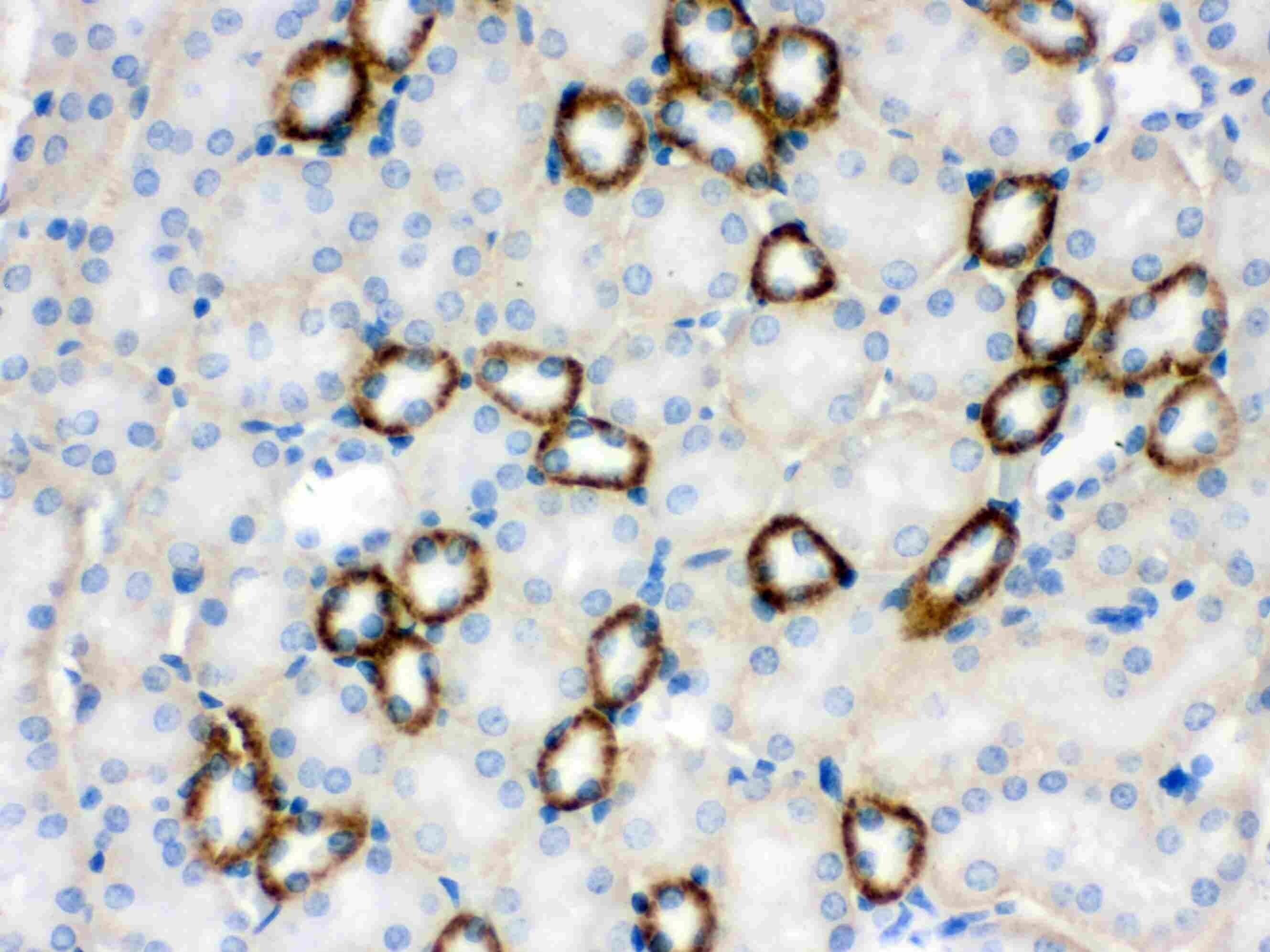
- Immunohistochemistry analysis of CYP1B1 in paraffin-embedded mouse kidney tissue. Antigen retrieval was performed on the tissue using citrate buffer (pH 6, 20 min) and blocked with 10% goat serum. Samples were incubated with CYP1B1 polyclonal antibody (Product # PA5-95277) at a 1 µg/mL dilution, followed by biotinylated goat anti-rabbit IgG (30 min, 37°C), and developed with Strepavidin-Biotin-Complex and DAB.
- Submitted by
- Invitrogen Antibodies (provider)
- Main image
- Experimental details
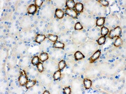
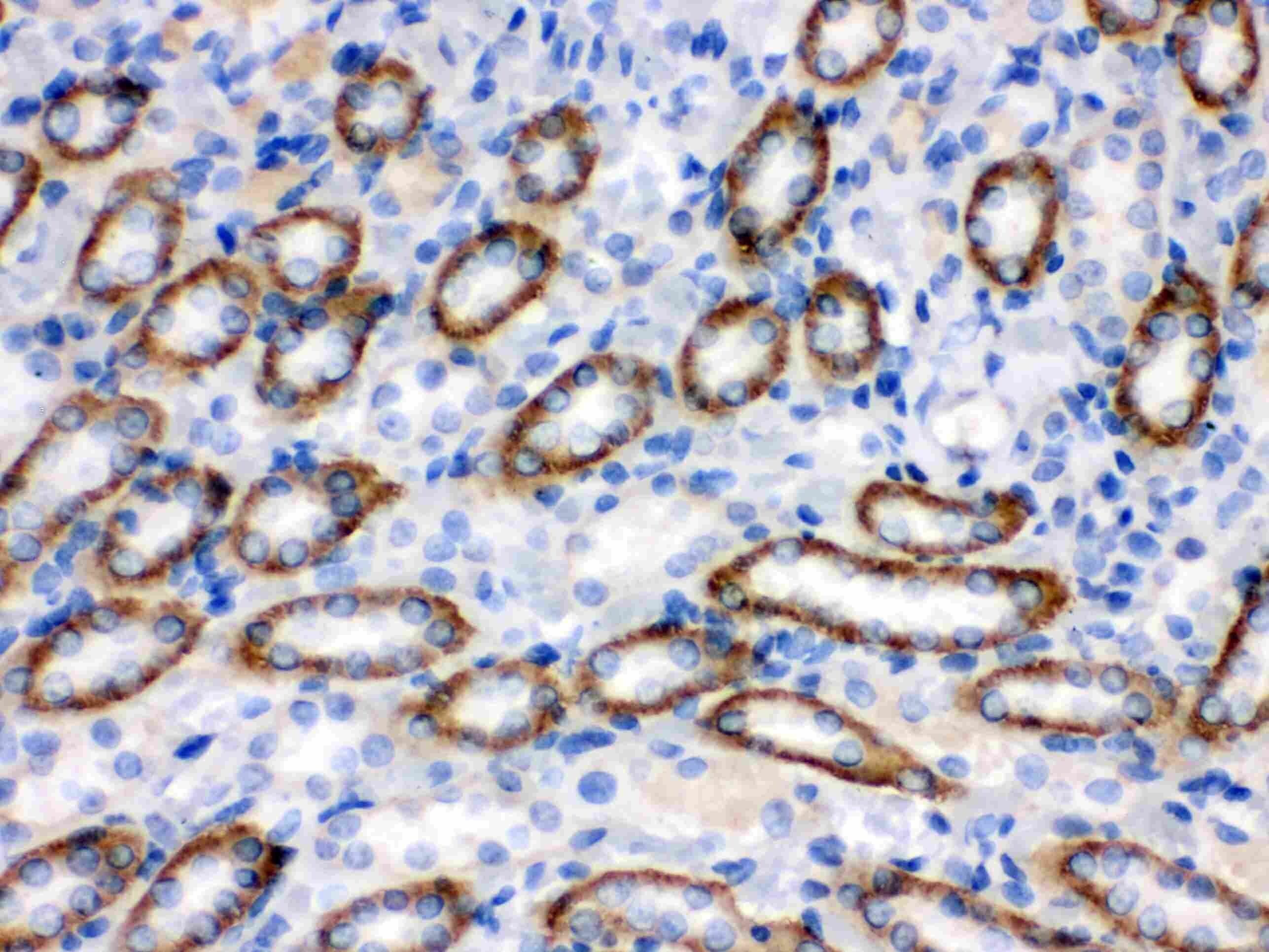
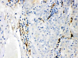
- Immunohistochemistry analysis of CYP1B1 in paraffin-embedded rat kidney tissue. Antigen retrieval was performed on the tissue using citrate buffer (pH 6, 20 min) and blocked with 10% goat serum. Samples were incubated with CYP1B1 polyclonal antibody (Product # PA5-95277) at a 1 µg/mL dilution, followed by biotinylated goat anti-rabbit IgG (30 min, 37°C), and developed with Strepavidin-Biotin-Complex and DAB.
- Submitted by
- Invitrogen Antibodies (provider)
- Main image
- Experimental details
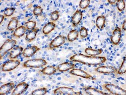
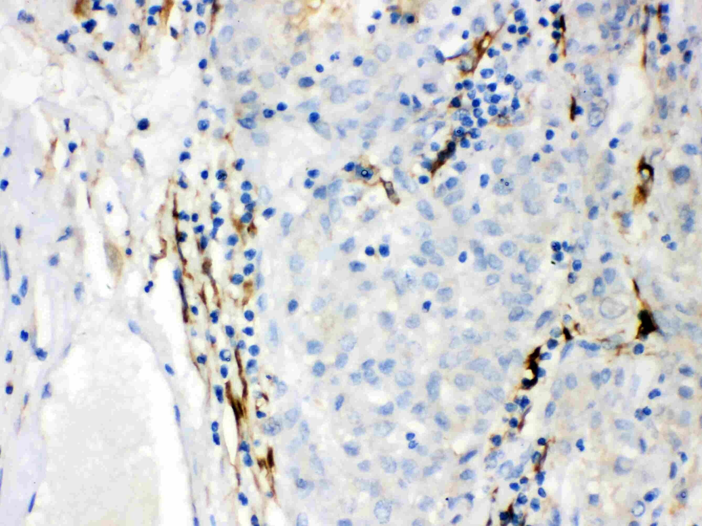
- Immunohistochemistry analysis of CYP1B1 in paraffin-embedded human liver cancer tissue. Antigen retrieval was performed on the tissue using citrate buffer (pH 6, 20 min) and blocked with 10% goat serum. Samples were incubated with CYP1B1 polyclonal antibody (Product # PA5-95277) at a 1 µg/mL dilution, followed by biotinylated goat anti-rabbit IgG (30 min, 37°C), and developed with Strepavidin-Biotin-Complex and DAB.
Supportive validation
- Submitted by
- Invitrogen Antibodies (provider)
- Main image
- Experimental details
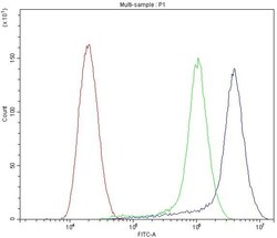
- Flow cytometry of CYP1B1 in SiHa cells (blue line), isotype control rabbit IgG (green line) and unlabeled (red line). Samples were blocked with 10% goat serum, incubated with CYP1B1 polyclonal antibody (Product # PA5-95277) at a dilution of 1 µg (per 1x10^6 cells), followed by 488 conjugated goat anti-rabbit IgG (30 min at 20°C) using a 5-10 µg (per 1x10^6 cells) dilution.
Supportive validation
- Submitted by
- Invitrogen Antibodies (provider)
- Main image
- Experimental details
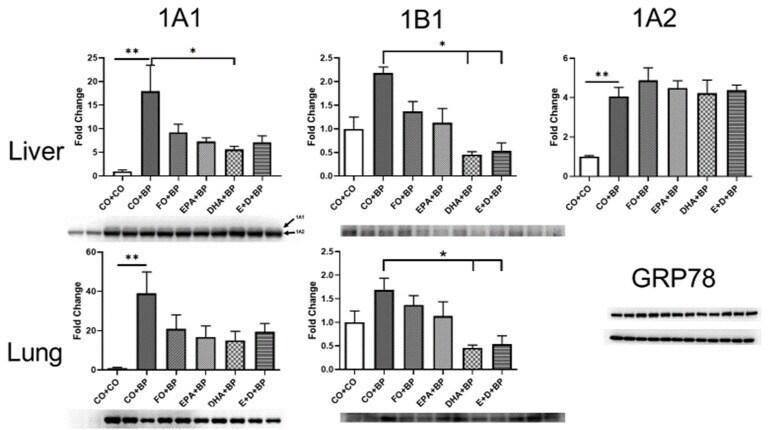
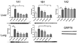
- Figure 7 Proteins isolated from liver and lung of mice, fed with CO, FO, EPA, DHA, EPA/DHA and treated with BP, were probed with Cyp1A1, 1B1 or 1A2 (liver only) antibodies by Western blot. FO, DHA and EPA + DHA significantly inhibited Cyp1b1 compared to CO group. Hepatic Cyp1A2 induced by BP treatment was also inhibited by FO, EPA, and DHA. *, p < 0.05, **, p < 0.01, ( n = 5).