 Explore
 Validate
 Learn
Learn Western blot
 Immunohistochemistry
Antibody data
- Antibody Data
- Antigen structure
- References [1]
- Comments [0]
- Validations
- Immunohistochemistry [1]
- Other assay [1]
Submit
Validation data
Reference
Comment
Report error
- Product number
- PA5-47469 - Provider product page

- Provider
- Invitrogen Antibodies
- Product name
- SEMA3E Polyclonal Antibody
- Antibody type
- Polyclonal
- Antigen
- Recombinant full-length protein
- Description
- In direct ELISAs, approximately 30% cross-reactivity with recombinant mouse (rm) Semaphorin 3E is observed and less than 10% cross-reactivity with rmSemaphorin 3A, rmSemaphorin 3B, rmSemaphorin 3C, rmSemaphorin 3F, and recombinant human (rh) Semaphorin 3A, rhSemaphorin 3C, rhSemaphorin 3D, and rhSemaphorin 3F is observed. Reconstitute at 0.2 mg/mL in sterile PBS.
- Reactivity
- Human, Mouse
- Host
- Goat
- Isotype
- IgG
- Vial size
- 100 µg
- Concentration
- 0.2 mg/mL
- Storage
- -20° C, Avoid Freeze/Thaw Cycles
Submitted references UHRF1 overexpression promotes osteosarcoma metastasis through altered exosome production and AMPK/SEMA3E suppression.
Wu SC, Kim A, Gu Y, Martinez DI, Zocchi L, Chen CC, Lopez J, Salcido K, Singh S, Wu J, Nael A, Benavente CA
Oncogenesis 2022 Sep 6;11(1):51
Oncogenesis 2022 Sep 6;11(1):51
No comments: Submit comment
Supportive validation
- Submitted by
- Invitrogen Antibodies (provider)
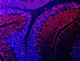
- Main image

- Experimental details
- Immunohistochemical analysis of SEMA3E in immersion fixed frozen sections of adult mouse brain. Samples were incubated in SEMA3E polyclonal antibody (Product # PA5-47469) using a dilution of 10 µg/mL overnight at 4 °C followed by NorthernLights™ 557-conjugated Anti-Goat IgG Secondary Antibody (red) and counterstained with DAPI (blue). Specific staining was localized to cerebellum.
Supportive validation
- Submitted by
- Invitrogen Antibodies (provider)
- Main image

- Experimental details
- Fig. 5 UHRF1 decreases SEMA3E expression through suppression of AMPK activation to induce angiogenesis. A , B Western blot analysis of ( A ) SEMA3E in SJSA-1 and SaOS-2 VC and KO osteosarcoma cell lines quantification of KO normalized to VC; and ( B ) activated AMPK (pAMPK) and total AMPK with the quantification of the ratio. UHRF1 used to confirm VC and KO status. beta-actin was used as a loading control. C Early phase sprouting angiogenesis (day 3) using human lung fibroblasts as assay control and SJSA-1 VC and KO cells with inducible sgRNA to knockout SEMA3E upon doxycycline addition in the fibrin gel bead assay. Quantified as the number of sprouts per bead. n = 30. ns not significant, * P < 0.05, **** P < 0.0001, by unpaired t test. D Model of UHRF1 oncogenic function in osteosarcoma. UHRF1 overexpression stimulates proliferation, exosome and uPA production that stimulates migration, invasion, and metastasis. UHRF1 also suppresses AMPK activation to inhibit SEMA3E and induce angiogenesis. Downstream of UHRF1, inhibitors of exosome secretion (e.g., GW4869), exosome endocytosis (CPZ), or uPA inhibitors (e.g., amiloride, BC11 hydrobromide) are attractive therapeutic options to decrease migration and metastasis. The development of UHRF1-targeted therapeutics might result in a beneficial decrease in both tumor growth and pulmonary metastases.
