Explore
 Validate
 Learn
Learn Western blot
Antibody data
- Antibody Data
- Antigen structure
- References [0]
- Comments [0]
- Validations
- Western blot [1]
- Immunohistochemistry [6]
- Flow cytometry [1]
Submit
Validation data
Reference
Comment
Report error
- Product number
- NBP2-03339 - Provider product page

- Provider
- Novus Biologicals
- Product name
- Mouse Monoclonal DUSP27/DUPD1 Antibody
- Antibody type
- Monoclonal
- Description
- Affinity purified.
- Reactivity
- Human
- Host
- Mouse
- Isotype
- IgG
- Vial size
- 0.1 ml
- Concentration
- 1.1 mg/ml
- Storage
- Store at -20C. Avoid freeze-thaw cycles.
No comments: Submit comment
Supportive validation
- Submitted by
- Novus Biologicals (provider)
- Main image

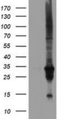
- Experimental details
- Western Blot: DUSP27/DUPD1 Antibody (7E4) [NBP2-03339] - DUSP27/DUPD1 Antibody (7E4) HEK293T cells were transfected with the pCMV6-ENTRY control (Left lane) or pCMV6-ENTRY DUSP27 (Right lane) cDNA for 48 hrs and lysed. Equivalent amounts of cell lysates (5 ug per lane) were separated by SDS-PAGE and immunoblotted with anti-DUSP27.
Supportive validation
- Submitted by
- Novus Biologicals (provider)
- Main image

- Experimental details
- Immunohistochemistry-Paraffin: DUSP27/DUPD1 Antibody (7E4) [NBP2-03339] - DUSP27/DUPD1 Antibody (7E4) Staining of paraffin-embedded Adenocarcinoma of Human endometrium tissue using anti-DUSP27 mouse monoclonal antibody.
- Submitted by
- Novus Biologicals (provider)
- Main image
- Experimental details
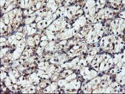
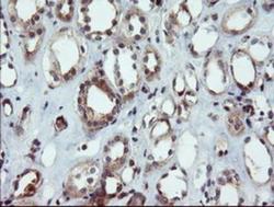
- Immunohistochemistry-Paraffin: DUSP27/DUPD1 Antibody (7E4) [NBP2-03339] - DUSP27/DUPD1 Antibody (7E4) Staining of paraffin-embedded Carcinoma of Human kidney tissue using anti-DUSP27 mouse monoclonal antibody.

- Submitted by
- Novus Biologicals (provider)
- Main image

- Experimental details
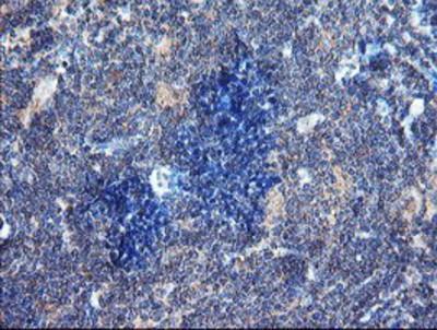
- Immunohistochemistry-Paraffin: DUSP27/DUPD1 Antibody (7E4) [NBP2-03339] - DUSP27/DUPD1 Antibody (7E4) Staining of paraffin-embedded Human lymphoma tissue using anti-DUSP27 mouse monoclonal antibody.
- Submitted by
- Novus Biologicals (provider)
- Main image
- Experimental details
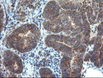
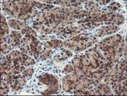
- Immunohistochemistry-Paraffin: DUSP27/DUPD1 Antibody (7E4) [NBP2-03339] - DUSP27/DUPD1 Antibody (7E4) Staining of paraffin-embedded Human pancreas tissue using anti-DUSP27 mouse monoclonal antibody.

- Submitted by
- Novus Biologicals (provider)
- Main image
- Experimental details
- Immunohistochemistry-Paraffin: DUSP27/DUPD1 Antibody (7E4) [NBP2-03339] - DUSP27/DUPD1 Antibody (7E4) Staining of paraffin-embedded Human Kidney tissue using anti-DUSP27 mouse monoclonal antibody.

- Submitted by
- Novus Biologicals (provider)
- Main image
- Experimental details
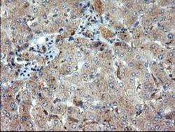
- Immunohistochemistry-Paraffin: DUSP27/DUPD1 Antibody (7E4) [NBP2-03339] - DUSP27/DUPD1 Antibody (7E4) Staining of paraffin-embedded Human liver tissue using anti-DUSP27 mouse monoclonal antibody.

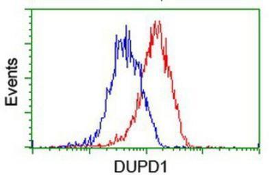
Supportive validation
- Submitted by
- Novus Biologicals (provider)
- Main image
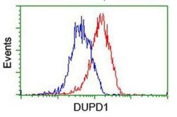
- Experimental details
- Flow Cytometry: DUSP27/DUPD1 Antibody (7E4) [NBP2-03339] - DUSP27/DUPD1 Antibody (7E4) Analysis of Hela cells, using anti-DUSP27 antibody, (Red), compared to a nonspecific negative control antibody (Blue).