 Explore
 Validate
 Learn
LearnNBP1-48337
antibody from Novus Biologicals
Targeting: CCR2
CC-CKR-2, CD192, CKR2, CMKBR2, FLJ78302, MCP-1-R
 Western blot
 Immunocytochemistry
Antibody data
- Antibody Data
- Antigen structure
- References [4]
- Comments [0]
- Validations
- Western blot [1]
- Immunohistochemistry [6]
- Flow cytometry [1]
Submit
Validation data
Reference
Comment
Report error
- Product number
- NBP1-48337 - Provider product page

- Provider
- Novus Biologicals
- Proper citation
- Novus Cat#NBP1-48337, RRID:AB_10011101
- Product name
- Rabbit Polyclonal CCR2 Antibody
- Antibody type
- Polyclonal
- Description
- Immunogen affinity purified.
- Reactivity
- Human, Rat, Bovine, Feline
- Host
- Rabbit
- Isotype
- IgG
- Vial size
- 0.1 ml
- Concentration
- 1 mg/ml
- Storage
- Store at -20C. Avoid freeze-thaw cycles.
Submitted references Expression of C-C motif chemokines and their receptors in bovine placentomes at spontaneous and induced parturition.
Direct role of the C-C motif chemokine receptor 2/monocyte chemoattractant protein 1 system in the feline cumulus oocyte complex†.
Kartogenin inhibits pain behavior, chondrocyte inflammation, and attenuates osteoarthritis progression in mice through induction of IL-10.
Possible Roles of CC- and CXC-Chemokines in Regulating Bovine Endometrial Function during Early Pregnancy.
Hirayama H, Sakumoto R, Koyama K, Yasuhara T, Hasegawa T, Inaba R, Fujii T, Naito A, Moriyasu S, Kageyama S
The Journal of reproduction and development 2020 Feb 14;66(1):49-55
The Journal of reproduction and development 2020 Feb 14;66(1):49-55
Direct role of the C-C motif chemokine receptor 2/monocyte chemoattractant protein 1 system in the feline cumulus oocyte complex†.
Rojo JL, Jaworski JP, Peluffo MC
Biology of reproduction 2019 Apr 1;100(4):1046-1056
Biology of reproduction 2019 Apr 1;100(4):1046-1056
Kartogenin inhibits pain behavior, chondrocyte inflammation, and attenuates osteoarthritis progression in mice through induction of IL-10.
Kwon JY, Lee SH, Na HS, Jung K, Choi J, Cho KH, Lee CY, Kim SJ, Park SH, Shin DY, Cho ML
Scientific reports 2018 Sep 14;8(1):13832
Scientific reports 2018 Sep 14;8(1):13832
Possible Roles of CC- and CXC-Chemokines in Regulating Bovine Endometrial Function during Early Pregnancy.
Sakumoto R, Hayashi KG, Fujii S, Kanahara H, Hosoe M, Furusawa T, Kizaki K
International journal of molecular sciences 2017 Mar 31;18(4)
International journal of molecular sciences 2017 Mar 31;18(4)
No comments: Submit comment
Supportive validation
- Submitted by
- Novus Biologicals (provider)
- Main image
- Experimental details
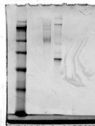
- Western Blot: CCR2 Antibody [NBP1-48337] - Analysis of CCR2 in rat lysate. Image courtesy of product review submitted by Rasha Elbaz.

Supportive validation
- Submitted by
- Novus Biologicals (provider)
- Main image

- Experimental details
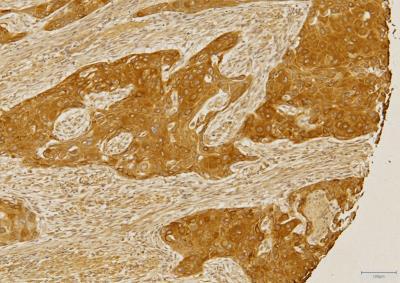
- Immunohistochemistry-Paraffin: CCR2 Antibody [NBP1-48337] - Analysis of FFPE section of human esophageal squamous cell carcinoma (SCC) using 5 ug/ml concentration of CCR2 antibody. Strong cytoplasmic-membranous immune-staining was observed in SCC cells and a relatively weaker staining was seen in the tumor stroma cells. [Magnification 10X]
- Submitted by
- Novus Biologicals (provider)
- Main image
- Experimental details
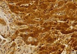
- Immunohistochemistry-Paraffin: CCR2 Antibody [NBP1-48337] - Analysis of FFPE tissue section of human liver cancer using 5 ug/ml concentration of CCR2 antibody. Intense cytoplasmic-membranous immune-staining was observed in the cancerous hepatocytes, whereas the staining was relatively weak in the cells adjacent to the fibrotic areas. [Magnification 40X]
- Submitted by
- Novus Biologicals (provider)
- Main image
- Experimental details
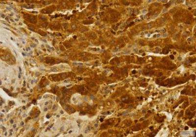
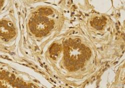
- Immunohistochemistry-Paraffin: CCR2 Antibody [NBP1-48337] - Analysis of FFPE tissue section of human normal breast using 5 ug/ml concentration of CCR2 antibody. The breast ductal/acinar epithelial cells showed a strong cytoplasmic-membranous CCR2 immune-positivity, whereas the intra-lobular connective tissue depicted weak staining. [Magnification 40X]

- Submitted by
- Novus Biologicals (provider)
- Main image

- Experimental details
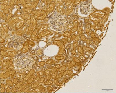
- Immunohistochemistry-Paraffin: CCR2 Antibody [NBP1-48337] - Analysis of FFPE tissue section of human normal kidney using 5 ug/ml concentration of CCR2 antibody. Strong cytoplasmic immunostaining was observed in the various renal cells (especially from tubules/ducts epithelium). The cells of Bowman's capsule depicted a very weak cytoplasmic staining for CCR2 protein. [Magnification 10X]
- Submitted by
- Novus Biologicals (provider)
- Main image

- Experimental details
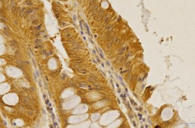
- Immunohistochemistry-Paraffin: CCR2 Antibody [NBP1-48337] - Analysis of FFPE tissue section of human rectal adenocarcinoma using 5 ug/ml concentration of CCR2 antibody. The cancerous cells in the section developed a uniform but specific CCR2 immunopositivity whereas a weak staining was seen in the tumor stroma cells. [Magnification 40X]
- Submitted by
- Novus Biologicals (provider)
- Main image
- Experimental details
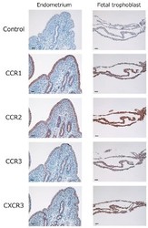
- Immunohistochemistry: CCR2 Antibody [NBP1-48337] - Localization of CCR1 (binds to CCL8, CCL14, and CCL16), CCR2 (binds to CCL2, CCL8, and CCL16), CCR3 (binds to CCL11), and CXCR3 (binds to CXCL10) in the bovine endometrium and fetal trophoblast obtained from cows in their 18th day of pregnancy. Intensive immunoreactivity was observed in endometrial epithelial cells, glandular epithelial cells, or fetal trophoblast. No positive immunoreactivity was observed in the negative control (Control). Scale bar = 50 um. Image collected and cropped by CiteAb from the following publication (http://www.mdpi.com/1422-0067/18/4/742), licensed under a CC-BY licence.

Supportive validation
- Submitted by
- Novus Biologicals (provider)
- Main image
- Experimental details
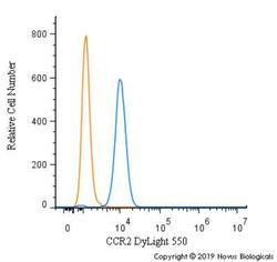
- Flow Cytometry: CCR2 Antibody [NBP1-48337] - An intracellular stain was performed on THP-1 cells with CCR Antibody NBP1-48337R (blue) and a matched isotype control (orange). Cells were fixed with 4% PFA and then permeabilized with 0.1% saponin. Cells were incubated in an antibody dilution of 5 ug/mL for 30 minutes at room temperature. Both antibodies were conjugated to DyLight 550.
