Explore
 Validate
 Learn
LearnNBP1-48338
antibody from Novus Biologicals
Targeting: CCR2
CC-CKR-2, CD192, CKR2, CMKBR2, FLJ78302, MCP-1-R
 Immunocytochemistry
 Flow cytometry
Antibody data
- Antibody Data
- Antigen structure
- References [4]
- Comments [0]
- Validations
- Flow cytometry [3]
Submit
Validation data
Reference
Comment
Report error
- Product number
- NBP1-48338 - Provider product page

- Provider
- Novus Biologicals
- Proper citation
- Novus Cat#NBP1-48338, RRID:AB_10011103
- Product name
- Rabbit Polyclonal CCR2 Antibody
- Antibody type
- Polyclonal
- Description
- Immunogen affinity purified.
- Reactivity
- Human, Mouse
- Host
- Rabbit
- Isotype
- IgG
- Vial size
- 0.1 ml
- Concentration
- 1 mg/ml
- Storage
- Store at -20C. Avoid freeze-thaw cycles.
Submitted references MMP12 Inhibits Corneal Neovascularization and Inflammation through Regulation of CCL2.
Neurodegeneration Enhances the Development of Arthritis.
Cardiac macrophages adopt profibrotic/M2 phenotype in infarcted hearts: Role of urokinase plasminogen activator.
Microenvironment-induced PTEN loss by exosomal microRNA primes brain metastasis outgrowth.
Wolf M, Clay SM, Zheng S, Pan P, Chan MF
Scientific reports 2019 Aug 9;9(1):11579
Scientific reports 2019 Aug 9;9(1):11579
Neurodegeneration Enhances the Development of Arthritis.
Lang SC, Harre U, Purohit P, Dietel K, Kienhöfer D, Hahn J, Baum W, Herrmann M, Schett G, Mielenz D
Journal of immunology (Baltimore, Md. : 1950) 2017 Mar 15;198(6):2394-2402
Journal of immunology (Baltimore, Md. : 1950) 2017 Mar 15;198(6):2394-2402
Cardiac macrophages adopt profibrotic/M2 phenotype in infarcted hearts: Role of urokinase plasminogen activator.
Carlson S, Helterline D, Asbe L, Dupras S, Minami E, Farris S, Stempien-Otero A
Journal of molecular and cellular cardiology 2017 Jul;108:42-49
Journal of molecular and cellular cardiology 2017 Jul;108:42-49
Microenvironment-induced PTEN loss by exosomal microRNA primes brain metastasis outgrowth.
Zhang L, Zhang S, Yao J, Lowery FJ, Zhang Q, Huang WC, Li P, Li M, Wang X, Zhang C, Wang H, Ellis K, Cheerathodi M, McCarty JH, Palmieri D, Saunus J, Lakhani S, Huang S, Sahin AA, Aldape KD, Steeg PS, Yu D
Nature 2015 Nov 5;527(7576):100-104
Nature 2015 Nov 5;527(7576):100-104
No comments: Submit comment
Supportive validation
- Submitted by
- Novus Biologicals (provider)
- Main image
- Experimental details
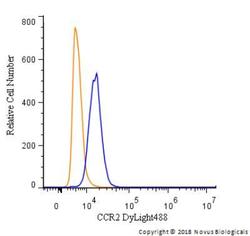
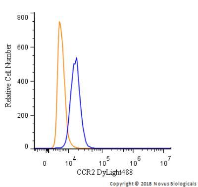
- Flow Cytometry: CCR2 Antibody [NBP1-48338] - An intracellular stain was performed on THP-1 cells with CCR2 Antibody NBP1-48338G (blue) and a matched isotype control (orange). Cells were fixed with 4% PFA and then permeabilized with 0.1% saponin. Cells were incubated in an antibody dilution of 2.5 ug/mL for 30 minutes at room temperature. Both antibodies were conjugated to DyLight 488.
- Submitted by
- Novus Biologicals (provider)
- Main image
- Experimental details
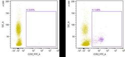
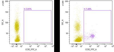
- Flow Cytometry: CCR2 Antibody [NBP1-48338] - The Alexa Fluor 488 conjugate of this antibody was used: Mouse peripheral blood mononuclear cells were unstained (left) or stained (right) with CCR2 antibody. Image from verified customer review.
- Submitted by
- Novus Biologicals (provider)
- Main image
- Experimental details
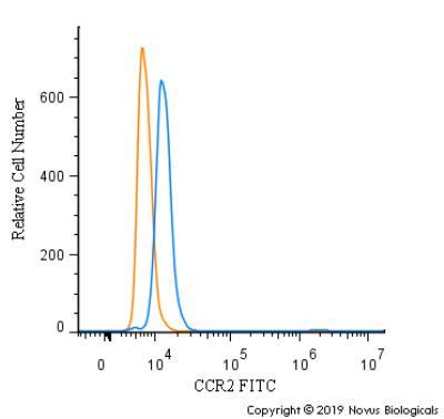
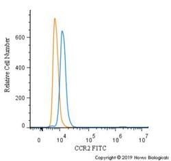
- Flow Cytometry: CCR2 Antibody [NBP1-48338] - An intracellular stain was performed on THP-1 cells with CCR2 Antibody NBP1-48338F (blue) and a matched isotype control (orange). Cells were fixed with 4% PFA and then permeabilized with 0.1% saponin. Cells were incubated in an antibody dilution of 10 ug/mL for 30 minutes at room temperature. Both antibodies were conjugated to FITC.