Explore
 Validate
 Learn
Learn Western blot
Antibody data
- Antibody Data
- Antigen structure
- References [1]
- Comments [0]
- Validations
- Western blot [2]
- Immunoprecipitation [1]
Submit
Validation data
Reference
Comment
Report error
- Product number
- MAB852 - Provider product page

- Provider
- R&D Systems
- Product name
- Human Bag-1 Antibody
- Antibody type
- Monoclonal
- Description
- Protein A or G purified from hybridoma culture supernatant. Detects the 36 kDa and 50 kDa isoforms of human Bag-1. Does not detect mouse Bag-1.
- Reactivity
- Human
- Host
- Mouse
- Conjugate
- Unconjugated
- Isotype
- IgG
- Antibody clone number
- GP3.10G3E2
- Vial size
- 100 ug
- Concentration
- LYOPH
- Storage
- Use a manual defrost freezer and avoid repeated freeze-thaw cycles. 12 months from date of receipt, -20 to -70 °C as supplied. 1 month, 2 to 8 °C under sterile conditions after reconstitution. 6 months, -20 to -70 °C under sterile conditions after reconstitution.
Submitted references Induction of apoptosis by Meretrix lusoria through reactive oxygen species production, glutathione depletion, and caspase activation in human leukemia cells.
Pan MH, Huang YT, Ho CT, Chang CI, Hsu PC, Sun Pan B
Life sciences 2006 Aug 15;79(12):1140-52
Life sciences 2006 Aug 15;79(12):1140-52
No comments: Submit comment
Supportive validation
- Submitted by
- R&D Systems (provider)
- Main image
- Experimental details
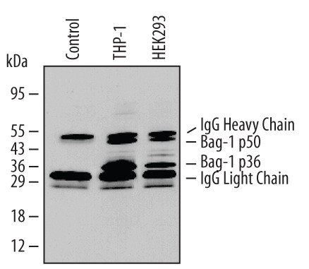
- Detection of Human Bag-1 by Western Blot. Western blot shows lysates of THP-1 human acute monocytic leukemia cell line and HEK293 human embryonic kidney cell line. PVDF Membrane was probed with 0.5 µg/mL of Mouse Anti-Human Bag-1 Monoclonal Antibody (Catalog # MAB852) followed by HRP-conjugated Anti-Mouse IgG Secondary Antibody (Catalog # HAF007). Specific bands were detected for Bag-1 at approximately 36 and 50 kDa (as indicated). This experiment was conducted under reducing conditions and using Immunoblot Buffer Group 6.

- Submitted by
- R&D Systems (provider)
- Main image
- Experimental details
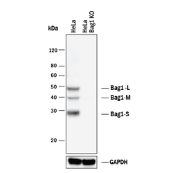
- Western Blot Shows Human Bag-1 Specificity by Using Knockout Cell Line. Western blot shows lysates of HeLa human cervical epithelial carcinoma parental cell line and Bag-1 knockout HeLa cell line (KO). PVDF membrane was probed with 0.5 µg/mL of Mouse Anti-Human Bag-1 Monoclonal Antibody (Catalog # MAB852) followed by HRP-conjugated Anti-Mouse IgG Secondary Antibody (Catalog # HAF018). Specific bands were detected for Bag-1 at approximately 29, 39, and 50 kDa (as indicated) in the parental HeLa cell line, but is not detectable in knockout HeLa cell line. GAPDH (Catalog # MAB5718) is shown as a loading control. This experiment was conducted under reducing conditions and using Immunoblot Buffer Group 1.

Supportive validation
- Submitted by
- R&D Systems (provider)
- Main image

- Experimental details
- Immunoprecipitation of Human Bag-1. Bag-1 was immunoprecipitated from lysates (5 x 10⁶ cells) of THP-1 human acute monocytic leukemia and HEK293 human embryonic kidney cell line following incubation with 3 µg Mouse Anti-Human Bag-1 Monoclonal Antibody (Catalog # MAB852) for 1-16 hours at 4 °C. Bag-1-antibody complexes were absorbed using Protein A Immunoprecipitin (Life Technologies). Immuno-precipitated Bag-1 (left panel) was detected by Western blot using 0.5 µg/mL Mouse CD30/TNFRSF8 Antigen Affinity-purified Polyclonal Antibody (Catalog # AF852). View our recommended buffer recipes for immunoprecipitation.