Explore
 Validate
 Learn
Learn Western blot
Antibody data
- Antibody Data
- Antigen structure
- References [1]
- Comments [0]
- Validations
- Western blot [2]
- Immunocytochemistry [1]
- Immunohistochemistry [5]
Submit
Validation data
Reference
Comment
Report error
- Product number
- HPA015269 - Provider product page

- Provider
- Atlas Antibodies
- Proper citation
- Atlas Antibodies Cat#HPA015269, RRID:AB_1857899
- Product name
- Anti-TES
- Antibody type
- Polyclonal
- Reactivity
- Human
- Host
- Rabbit
- Conjugate
- Unconjugated
- Antigen sequence
QLPAHDQDPSKCHELSPREVKEMEQFVKKYKSEAL
GVGDVKLPCEMDAQGPKQMNIPGGDRSTPAAVGAM
EDKSAEHKRTQYSCYCCKLSMKEGDPAIYAERAGY
DKLWHPACFVCSTCHELLVD- Isotype
- IgG
- Vial size
- 100 µl
- Storage
- Store at +4°C for short term storage. Long time storage is recommended at -20°C.
Submitted references RNA Deep Sequencing as a Tool for Selection of Cell Lines for Systematic Subcellular Localization of All Human Proteins
Danielsson F, Wiking M, Mahdessian D, Skogs M, Ait Blal H, Hjelmare M, Stadler C, Uhlén M, Lundberg E
Journal of Proteome Research 2013 January;12(1):299-307
Journal of Proteome Research 2013 January;12(1):299-307
No comments: Submit comment
Supportive validation
Supportive validation
- Submitted by
- Atlas Antibodies (provider)
- Enhanced method
- Orthogonal validation
- Main image

- Experimental details
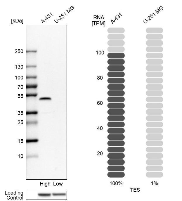
- Western blot analysis in human cell lines A-431 and U-251MG using Anti-TES antibody. Corresponding TES RNA-seq data are presented for the same cell lines. Loading control: Anti-HSP90B1.
Supportive validation
- Submitted by
- Atlas Antibodies (provider)
- Main image
- Experimental details
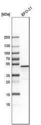
- Western blot analysis in human cell line EFO-21.
- Sample type
- HUMAN

Supportive validation
- Submitted by
- Atlas Antibodies (provider)
- Main image

- Experimental details
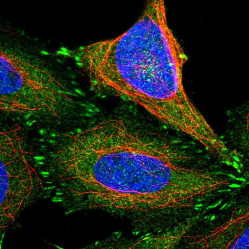
- Immunofluorescent staining of human cell line SiHa shows localization to cytosol & focal adhesion sites.
- Sample type
- HUMAN
Supportive validation
- Submitted by
- Atlas Antibodies (provider)
- Main image
- Experimental details
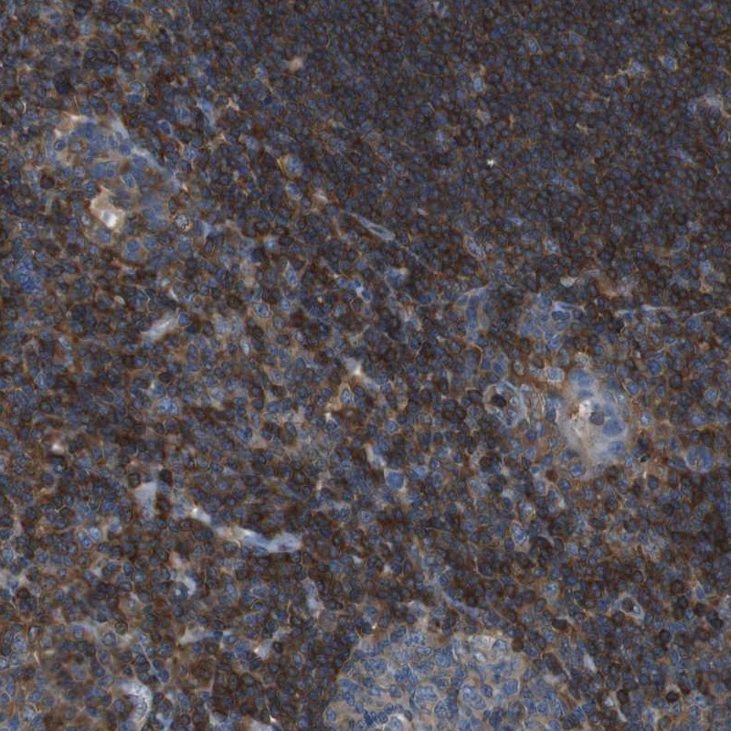
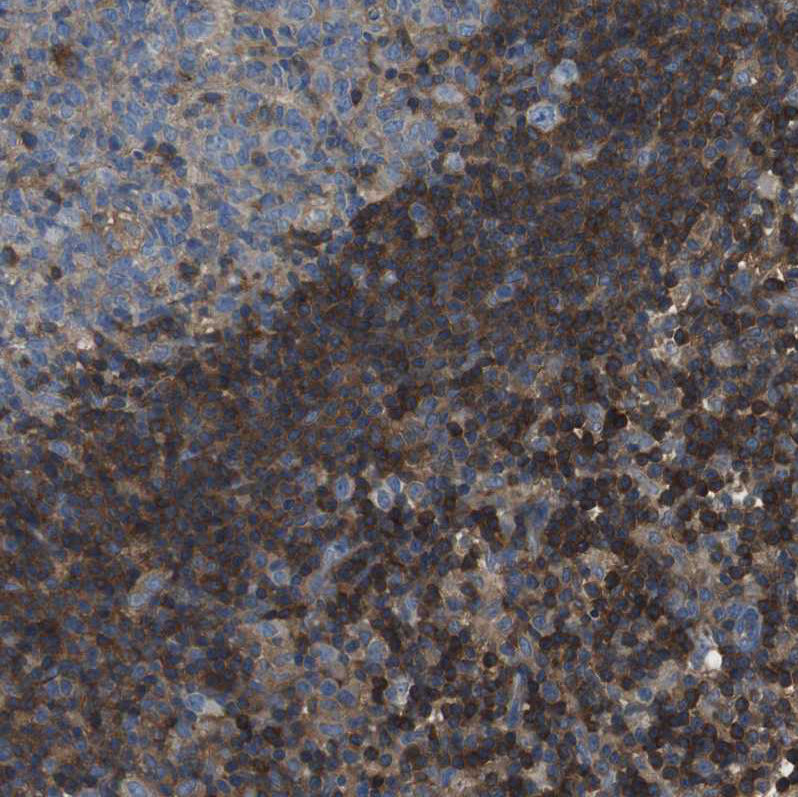
- Immunohistochemical staining of human tonsil shows strong cytoplasmic positivity in lymphoid cells outside reaction centra.
- Sample type
- HUMAN
- Submitted by
- Atlas Antibodies (provider)
- Main image
- Experimental details
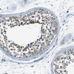
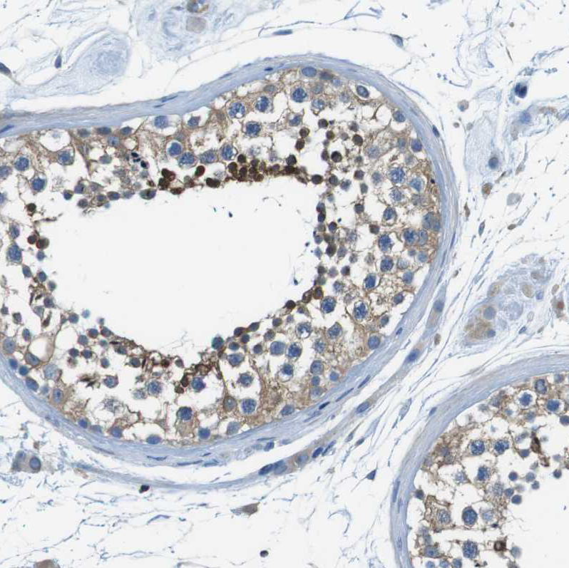
- Immunohistochemical staining of human testis shows moderate cytoplasmic positivity in cells in seminiferous ducts.
- Sample type
- HUMAN
- Submitted by
- Atlas Antibodies (provider)
- Main image
- Experimental details
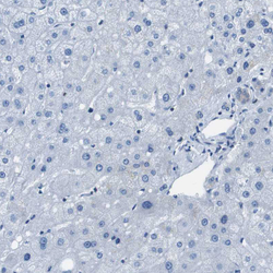
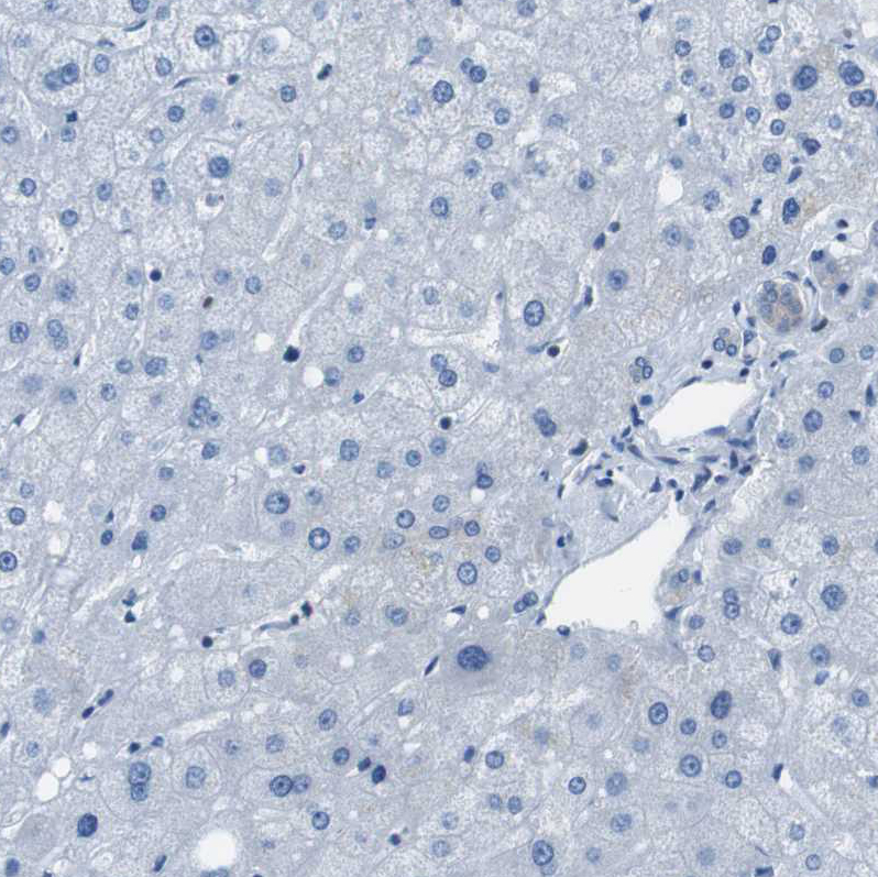
- Immunohistochemical staining of human liver shows no cytoplasmic positivity in hepatocytes as expected.
- Sample type
- HUMAN
- Submitted by
- Atlas Antibodies (provider)
- Main image
- Experimental details
- Immunohistochemical staining of human lymphoid tissues shows moderate cytoplasmic positivity in non-germinal center cells.
- Sample type
- HUMAN
- Submitted by
- Atlas Antibodies (provider)
- Main image

- Experimental details
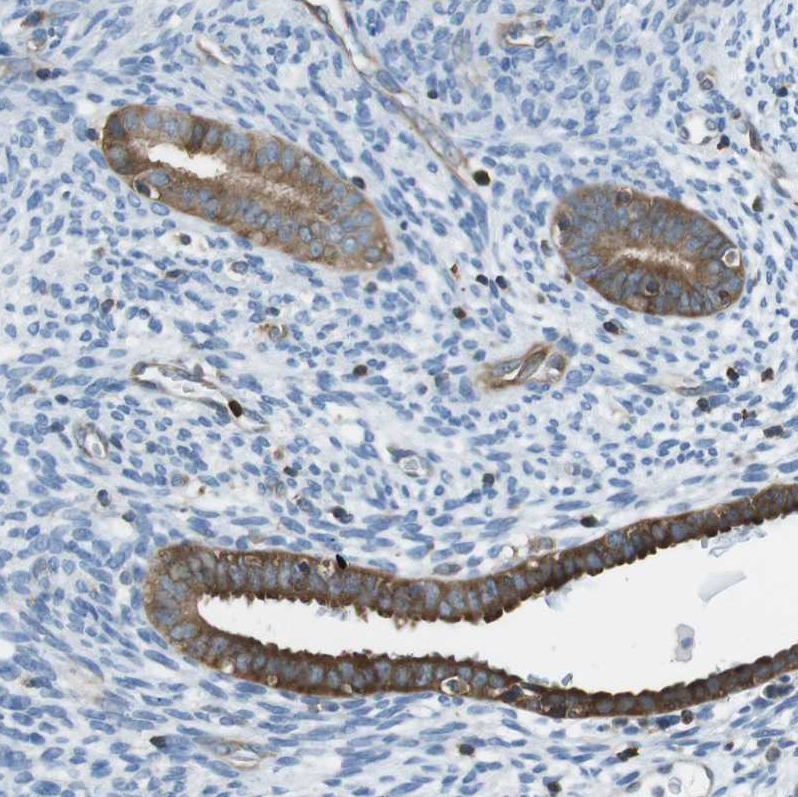
- Immunohistochemical staining of human endometrium moderate cytoplasmic positivity in glandular cells.
- Sample type
- HUMAN