 Explore
 Validate
 Learn
Learn Western blot
Antibody data
- Antibody Data
- Antigen structure
- References [0]
- Comments [0]
- Validations
- Western blot [3]
- Immunocytochemistry [1]
- Immunohistochemistry [1]
Submit
Validation data
Reference
Comment
Report error
- Product number
- HPA018123 - Provider product page

- Provider
- Atlas Antibodies
- Proper citation
- Atlas Antibodies Cat#HPA018123, RRID:AB_1857900
- Product name
- Anti-TES
- Antibody type
- Polyclonal
- Reactivity
- Human
- Host
- Rabbit
- Conjugate
- Unconjugated
- Antigen sequence
YCDSEKPRCAGCDELIFSNEYTQAENQNWHLKHFC
CFDCDSILAGEIYVMVNDKPVCKPCYVKNHAVVCQ
GCHNAIDPEVQRVTYNNFSWHASTECFLCSCCSKC
LIGQKFMPVEGMVFCSVECKK- Isotype
- IgG
- Vial size
- 100 µl
- Storage
- Store at +4°C for short term storage. Long time storage is recommended at -20°C.
No comments: Submit comment
Supportive validation
Supportive validation
Supportive validation
- Submitted by
- Atlas Antibodies (provider)
- Enhanced method
- Orthogonal validation
- Main image

- Experimental details
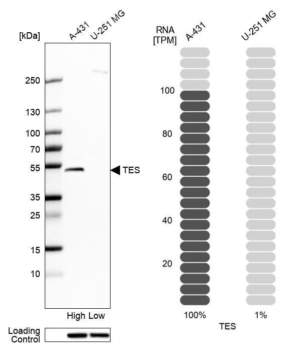
- Western blot analysis in human cell lines A-431 and U-251MG using Anti-TES antibody. Corresponding TES RNA-seq data are presented for the same cell lines. Loading control: Anti-HSP90B1.
Supportive validation
- Submitted by
- Atlas Antibodies (provider)
- Enhanced method
- Independent antibody validation
- Main image
- Experimental details
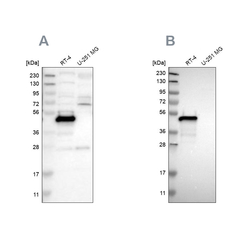
- Western blot analysis using Anti-TES antibody HPA018123 (A) shows similar pattern to independent antibody HPA015269 (B).

Supportive validation
- Submitted by
- Atlas Antibodies (provider)
- Main image

- Experimental details
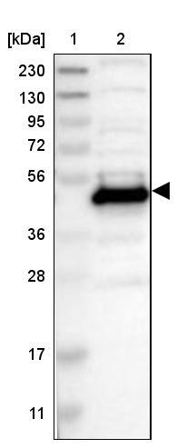
- Lane 1: Marker [kDa] 230, 130, 95, 72, 56, 36, 28, 17, 11Lane 2: Human cell line RT-4
Supportive validation
- Submitted by
- Atlas Antibodies (provider)
- Main image
- Experimental details
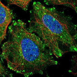
- Immunofluorescent staining of human cell line SiHa shows localization to plasma membrane, cytosol & focal adhesion sites.
- Sample type
- HUMAN

Supportive validation
- Submitted by
- Atlas Antibodies (provider)
- Main image
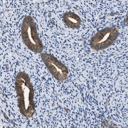
- Experimental details
- Immunohistochemical staining of human endometrium shows moderate cytoplasmic positivity in glandular cells.
- Sample type
- HUMAN
