 Explore
 Validate
 Learn
Learn Western blot
Antibody data
- Antibody Data
- Antigen structure
- References [3]
- Comments [0]
- Validations
- Western blot [2]
- Immunocytochemistry [3]
- Immunohistochemistry [1]
- Flow cytometry [1]
- Other assay [1]
Submit
Validation data
Reference
Comment
Report error
- Product number
- 45-5900 - Provider product page

- Provider
- Invitrogen Antibodies
- Product name
- Cyclophilin F Monoclonal Antibody (E11AE12BD4)
- Antibody type
- Monoclonal
- Antigen
- Recombinant full-length protein
- Description
- Positive controls: Isolated mitochondria from Human heart, Bovine heart, Rat heart, Mouse heart, HepG2 cells; Cultured Human embryonic lung-derived fibroblasts (strain MRC5); Human cerebellum tissue; HL60 cells.
- Reactivity
- Human, Mouse, Rat, Bovine
- Host
- Mouse
- Isotype
- IgG
- Antibody clone number
- E11AE12BD4
- Vial size
- 100 µg
- Concentration
- 1 mg/mL
- Storage
- 4° C, do not freeze
Submitted references Inner mitochondrial membrane protein MPV17 mutant mice display increased myocardial injury after ischemia/reperfusion.
Deficiency of miR-208a Exacerbates CCl(4)-Induced Acute Liver Injury in Mice by Activating Cell Death Pathways.
Liproxstatin-1 protects the mouse myocardium against ischemia/reperfusion injury by decreasing VDAC1 levels and restoring GPX4 levels.
Madungwe NB, Feng Y, Imam Aliagan A, Tombo N, Kaya F, Bopassa JC
American journal of translational research 2020;12(7):3412-3428
American journal of translational research 2020;12(7):3412-3428
Deficiency of miR-208a Exacerbates CCl(4)-Induced Acute Liver Injury in Mice by Activating Cell Death Pathways.
Bala S, Calenda CD, Catalano D, Babuta M, Kodys K, Nasser IA, Vidal B, Szabo G
Hepatology communications 2020 Oct;4(10):1487-1501
Hepatology communications 2020 Oct;4(10):1487-1501
Liproxstatin-1 protects the mouse myocardium against ischemia/reperfusion injury by decreasing VDAC1 levels and restoring GPX4 levels.
Feng Y, Madungwe NB, Imam Aliagan AD, Tombo N, Bopassa JC
Biochemical and biophysical research communications 2019 Dec 10;520(3):606-611
Biochemical and biophysical research communications 2019 Dec 10;520(3):606-611
No comments: Submit comment
Supportive validation
- Submitted by
- Invitrogen Antibodies (provider)
- Main image
- Experimental details
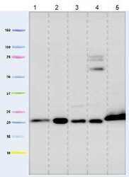
- Western blot analysis of Cyclophilin F in isolated mitochondria using a Cyclophilin F Monoclonal antibody (Product # 45-5900) at a concentration of 1 µg/mL. Lane 1: Isolated mitochondria from Human heart, 5 µg loaded. Lane 2: Isolated mitochondria from Bovine heart, 1 µg loaded. Lane 3: Isolated mitochondria from Rat heart, 10 µg loaded. Lane 4: Isolated mitochondria from Mouse heart, 10 µg loaded. Lane 5: Isolated mitochondria from HepG2 cells, 20 µg loaded.

- Submitted by
- Invitrogen Antibodies (provider)
- Main image

- Experimental details
- Western blot analysis was performed on membrane enriched cell extracts (30 µg lysate) of PC-12 (Lane 1), Jurkat (Lane 2), HT-29 (Lane 3), DU-145 (Lane 4), PANC-1 (Lane 5), U-937 (Lane 6), HEK-293 (Lane 7) and tissue extract of Rat Heart (Lane 8). The blot was probed with Anti- Cyclophilin F Monoclonal Antibody (Product # 45-5900, 1 µg/ml) and detected by chemiluminescence using Goat anti-Mouse IgG (H+L) Superclonal™ Secondary Antibody, HRP conjugate (Product # A28177, 0.25µg/ml, 1:4000 dilution). A 22 kDa band corresponding to Cyclophilin F was observed across the cell lines and tissue tested.

Supportive validation
- Submitted by
- Invitrogen Antibodies (provider)
- Main image

- Experimental details
- Immunofluorescent analysis of Cyclophilin F in MRC5 cells using a Cyclophilin F Monoclonal antibody (Product # 45-5900) at a concentration of 1 µg/mL. Cultured Human embryonic lung-derived fibroblasts (strain MRC5), were fixed, treated for heat-induced antigen retrieval, and permeabilized. Detection was perfomed by an AlexaFluor® 488-conjugated-goat-anti-mouse IgG1 isotype specific secondary antibody (2 µg/mL).

- Submitted by
- Invitrogen Antibodies (provider)
- Main image

- Experimental details
- Immunofluorescence analysis of Cyclophilin F in HEK293 cells using a Cyclophilin F Monoclonal antibody (Product # 45-5900) at a dilution of 1:200. HEK293 cells were fixed with paraformaldehyde, permeabilized with 1% triton X-100, and blocked with 10% goat serum for 1 hour at room temperature. Detection was perfomed by a goat anti-mouse IgG secondary antibody at a 1:300 dilution.

Supportive validation
- Submitted by
- Invitrogen Antibodies (provider)
- Main image

- Experimental details
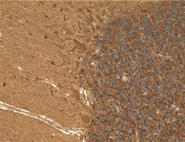
- Immunohistochemical analysis of Cyclophilin F in Human cerebellum using a Cyclophilin F Monoclonal antibody (Product # 45-5900) at a concentration of 1 µg/mL. Human cerebellum tissue was formalin fixed and paraffin embedded. Note: immunoactivity is most intense in neuronal cell bodies, most notably in the large Purkinje cells.
Supportive validation
- Submitted by
- Invitrogen Antibodies (provider)
- Main image

- Experimental details
- Flow cytometric analysis of Cyclophilin F in HL60 cells using an Cyclophilin F monoclonal antibody (Product # 45-5900) at 1 µg/mL is depicted by the blue line. The red line indicates an isotype control antibody.

Supportive validation
- Submitted by
- Invitrogen Antibodies (provider)
- Main image

- Experimental details
- 4 FIG. Increased expression of genes involved in necrosis in miR-208a KO mice after acute CCl 4 treatment. WT or miR-208a KO mice received oil (n = 4-5/group) or CCl 4 (n = 6/group) treatment for 48 hours. Whole-liver cell lysates were used to determine the protein levels of Bax (A), CypD (B), PCNA (C), and cyclin D1 (D) by western blot analysis. beta-actin was used as a loading control. Density units are shown in the bar diagram from mice, Bax (n = 4 [oil], n = 6 [CCl 4 ]), CypD (n = 4 [oil]; n = 6 [CCl 4 ]), PCNA (n = 3 [oil], n = 3 [CCl 4 ]), and cyclin D1 (n = 3 [oil], n = 3 [CCl 4 ]). * P < 0.05 versus oil-treated WT mice, # P < 0.05 versus CCl 4 -treated WT mice.
