Explore
 Validate
 Learn
Learn Western blot
Antibody data
- Antibody Data
- Antigen structure
- References [4]
- Comments [0]
- Validations
- Western blot [3]
- Immunohistochemistry [1]
- Other assay [2]
Submit
Validation data
Reference
Comment
Report error
- Product number
- 41-5800 - Provider product page

- Provider
- Invitrogen Antibodies
- Product name
- Connexin 45 Monoclonal Antibody (5C7G1)
- Antibody type
- Monoclonal
- Antigen
- Synthetic peptide
- Description
- 41-5800 was used in the western blot analysis to successfully detect Connexin 45 in mouse atrium homogenate.
- Reactivity
- Human, Mouse
- Host
- Mouse
- Isotype
- IgG
- Antibody clone number
- 5C7G1
- Vial size
- 100 µg
- Concentration
- 0.5 mg/mL
- Storage
- -20°C
Submitted references An offset ON-OFF receptive field is created by gap junctions between distinct types of retinal ganglion cells.
A Self-Regulating Gap Junction Network of Amacrine Cells Controls Nitric Oxide Release in the Retina.
Mitochondrial connexin40 regulates mitochondrial calcium uptake in coronary endothelial cells.
Developmentally dynamic colocalization patterns of DSCAM with adhesion and synaptic proteins in the mouse retina.
Cooler S, Schwartz GW
Nature neuroscience 2021 Jan;24(1):105-115
Nature neuroscience 2021 Jan;24(1):105-115
A Self-Regulating Gap Junction Network of Amacrine Cells Controls Nitric Oxide Release in the Retina.
Jacoby J, Nath A, Jessen ZF, Schwartz GW
Neuron 2018 Dec 5;100(5):1149-1162.e5
Neuron 2018 Dec 5;100(5):1149-1162.e5
Mitochondrial connexin40 regulates mitochondrial calcium uptake in coronary endothelial cells.
Guo R, Si R, Scott BT, Makino A
American journal of physiology. Cell physiology 2017 Apr 1;312(4):C398-C406
American journal of physiology. Cell physiology 2017 Apr 1;312(4):C398-C406
Developmentally dynamic colocalization patterns of DSCAM with adhesion and synaptic proteins in the mouse retina.
de Andrade GB, Kunzelman L, Merrill MM, Fuerst PG
Molecular vision 2014;20:1422-33
Molecular vision 2014;20:1422-33
No comments: Submit comment
Supportive validation
- Submitted by
- Invitrogen Antibodies (provider)
- Main image

- Experimental details
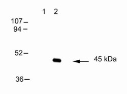
- Western blot analysis of lysates prepared from non-transfected (lane 1) and connexin 45-transfected (lane 2) HeLa cells using mouse anti-connexin 45 antibody (Product # 41-5800).

- Submitted by
- Invitrogen Antibodies (provider)
- Main image

- Experimental details
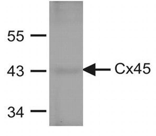
- Western blot analysis of Connexin45 (Cx45) was performed by loading 20 µg of protein from mouse atrium homogenate (not boiled) and 7 µL of EZ-RUN Prestained Rec Protein Ladder per well onto a 10% Tris-Hcl polyacrylamide gel. Proteins were transferred to a PVDF membrane nd blocked in blocking buffer composed of 20 mM Tris-HCl pH 8, 150 mM NaCl and 0.2% Tween-20 (TBST) supplemented with 5% skim milk powder (Carnation) for 1 hr at room temperature. Cx45 was detected at ~45 kDa using an anti-Cx45 mouse monoclonal antibody (Product # 41-5800) at a dilution of 1 µg/mL in TBST supplemented with 1% skim milk powder and incubated overnight at 4C on a rocking platform, followed by washing and incubation with a goat anti-mouse IgG horse radish peroxidase (HRP) conjugated secondary antibody at a dilution of 1:5,000 in TBST for 1 hr at room temperature. Chemiluminescent detection was performed using Pierce ECL Western blotting substrate (Product # 32209). Data courtesy of Dr. James Nagy's lab.
- Submitted by
- Invitrogen Antibodies (provider)
- Main image
- Experimental details
- Western blot analysis of lysates prepared from non-transfected (lane 1) and connexin 45-transfected (lane 2) HeLa cells using mouse anti-connexin 45 antibody (Product # 41-5800).

Supportive validation
- Submitted by
- Invitrogen Antibodies (provider)
- Main image
- Experimental details
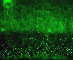
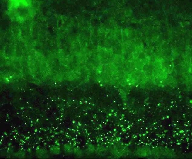
- Immunohistochemical analysis of gap junctions in frozen inner plexiform layer of mouse retina using mouse anti-connexin 45 antibody (Product # 41-5800). Image is provided courtesy of Dr. James Nagy, University of Manitoba, Canada.
Supportive validation
- Submitted by
- Invitrogen Antibodies (provider)
- Main image

- Experimental details
- NULL
- Submitted by
- Invitrogen Antibodies (provider)
- Main image

- Experimental details
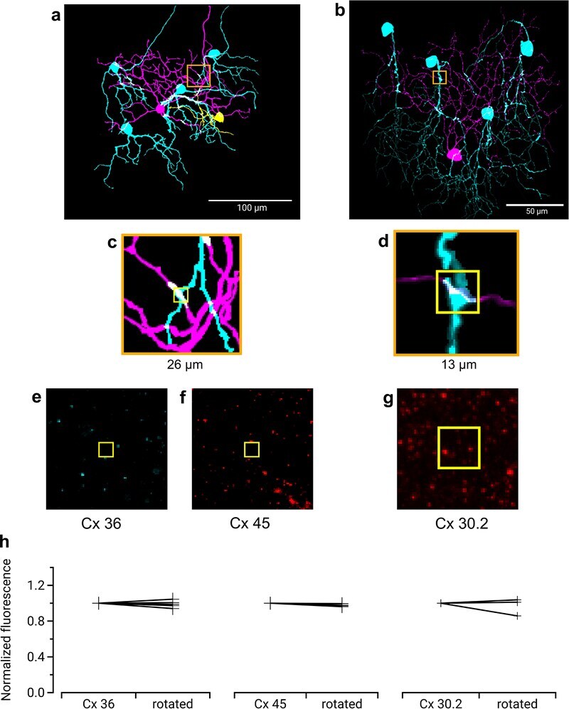
- Extended Data Fig. 4 Immunohistochemistry for three types of Connexin at RGC contact points shows negative results Three connexins were evaluated for presence at the regions of contact between an F-mini-ON and multiple F-mini-OFF RGCs, n = 1 of each experiment. a,b, Full depth maximum intensity projection images of a Neurobiotin-filled F-mini-ON RGC (magenta),the connected F-mini-OFF RGCs (cyan), and a cell of unclassified type due to insufficiently filled dendrites (yellow). Tracing, segmentation, and masking were performed manually. Image brightness was scaled separately by cell type for illustration here but not for analysis. c,d Thin projection images of regions in orange squares in a,b showing an example RGC crossing point with yellow square for spatial reference. Stack depth is 3.5 mum. e-g, The same region and depth as in c,d, showing the IHC channels for the three connexin proteins. h, Quantification of overlap between connexin images and RGC contact region masks. Values are similar before and after a 90 degree rotation of the connexin image. Points mark the overlap of the single F-mini-ON RGC with each F-mini-OFF RGC in the image.