Antibody data
- Antibody Data
- Antigen structure
- References [6]
- Comments [0]
- Validations
- Western blot [2]
- Immunocytochemistry [2]
- Immunohistochemistry [2]
- Flow cytometry [1]
Submit
Validation data
Reference
Comment
Report error
- Product number
- MA5-13212 - Provider product page

- Provider
- Invitrogen Antibodies
- Product name
- CD79a Monoclonal Antibody (HM47/A9)
- Antibody type
- Monoclonal
- Antigen
- Synthetic peptide
- Description
- MA5-13212 targets CD79a in IF, IHC (P), and WB applications and shows reactivity with Bovine, Equine, Guinea Pig, Human, mouse, Non-human primate, Porcine, Rabbit, Chicken, and Rat samples. The MA5-13212 immunogen is a synthetic peptide consisting of GTYQDVGSLNIADVQ of human CD79a protein. MA5-13212 is not functional in IHC (P) in its B format.
- Reactivity
- Human, Mouse, Rat, Bovine, Chicken/Avian, Guinea Pig, Porcine, Rabbit
- Host
- Mouse
- Isotype
- IgG
- Antibody clone number
- HM47/A9
- Vial size
- 500 µL
- Concentration
- 400 mg/mL
- Storage
- 4° C
Submitted references Lymphocytic leukemia in a captive dhole (Cuon alpinus).
Blastic plasmacytoid dendritic cell neoplasm with leukemic presentation: an Italian multicenter study.
Rats deficient for p53 are susceptible to spontaneous and carcinogen-induced tumorigenesis.
Pneumocystis mediates overexpression of antizyme inhibitor resulting in increased polyamine levels and apoptosis in alveolar macrophages.
A tumor-suppressor function for NFATc3 in T-cell lymphomagenesis by murine leukemia virus.
Immunohistochemical and histochemical stains for differentiating canine cutaneous round cell tumors.
Scala C, Ortiz K, Nicolier A, Briend-Marchal A
Journal of zoo and wildlife medicine : official publication of the American Association of Zoo Veterinarians 2013 Mar;44(1):204-7
Journal of zoo and wildlife medicine : official publication of the American Association of Zoo Veterinarians 2013 Mar;44(1):204-7
Blastic plasmacytoid dendritic cell neoplasm with leukemic presentation: an Italian multicenter study.
Pagano L, Valentini CG, Pulsoni A, Fisogni S, Carluccio P, Mannelli F, Lunghi M, Pica G, Onida F, Cattaneo C, Piccaluga PP, Di Bona E, Todisco E, Musto P, Spadea A, D'Arco A, Pileri S, Leone G, Amadori S, Facchetti F, GIMEMA-ALWP (Gruppo Italiano Malattie EMatologiche dell'Adulto, Acute Leukemia Working Party)
Haematologica 2013 Feb;98(2):239-46
Haematologica 2013 Feb;98(2):239-46
Rats deficient for p53 are susceptible to spontaneous and carcinogen-induced tumorigenesis.
Yan HX, Wu HP, Ashton C, Tong C, Ying QL
Carcinogenesis 2012 Oct;33(10):2001-5
Carcinogenesis 2012 Oct;33(10):2001-5
Pneumocystis mediates overexpression of antizyme inhibitor resulting in increased polyamine levels and apoptosis in alveolar macrophages.
Liao CP, Lasbury ME, Wang SH, Zhang C, Durant PJ, Murakami Y, Matsufuji S, Lee CH
The Journal of biological chemistry 2009 Mar 20;284(12):8174-84
The Journal of biological chemistry 2009 Mar 20;284(12):8174-84
A tumor-suppressor function for NFATc3 in T-cell lymphomagenesis by murine leukemia virus.
Glud SZ, Sørensen AB, Andrulis M, Wang B, Kondo E, Jessen R, Krenacs L, Stelkovics E, Wabl M, Serfling E, Palmetshofer A, Pedersen FS
Blood 2005 Nov 15;106(10):3546-52
Blood 2005 Nov 15;106(10):3546-52
Immunohistochemical and histochemical stains for differentiating canine cutaneous round cell tumors.
Fernandez NJ, West KH, Jackson ML, Kidney BA
Veterinary pathology 2005 Jul;42(4):437-45
Veterinary pathology 2005 Jul;42(4):437-45
No comments: Submit comment
Supportive validation
- Submitted by
- Invitrogen Antibodies (provider)
- Main image

- Experimental details
- Western blot of CD79a using CD79a Monoclonal Antibody (Product # MA5-13212) on Raji Cells.
- Submitted by
- Invitrogen Antibodies (provider)
- Main image

- Experimental details
- Western blot was performed using Anti-CD79a Monoclonal Antibody (HM47/A9) (Product # MA5-13212) and a 40kDa band corresponding to CD79a was observed across cell lines tested. Whole cell extracts (30 µg lysate) of Raji (Lane 1), Ramos (Lane 2), Daudi (Lane 3), HL-60 (Lane 4), SH-SY5Y (Lane 5), NTERA-2 cl.D1 (Lane 6) were electrophoresed using NuPAGE™ 12% Bis-Tris Protein Gel (Product # NP0342BOX). Resolved proteins were then transferred onto a Nitrocellulose membrane (Product # IB23001) by iBlot® 2 Dry Blotting System (Product # IB21001). The blot was probed with the primary antibody (1:50) and detected by chemiluminescence with Goat anti-Mouse IgG (H+L) Superclonal™ Recombinant Secondary Antibody, HRP (Product # A28177,1:5000) using the iBright FL 1000 (Product # A32752). Chemiluminescent detection was performed using Novex® ECL Chemiluminescent Substrate Reagent Kit (Product # WP20005).
Supportive validation
- Submitted by
- Invitrogen Antibodies (provider)
- Main image

- Experimental details
- Immunofluorescence analysis of CD79a was performed using 70% confluent log phase K-562 cells. The cells were fixed with 4% paraformaldehyde for 10 minutes, permeabilized with 0.1% Triton™ X-100 for 10 minutes, and blocked with 1% BSA for 1 hour at room temperature. The cells were labeled with CD79a Mouse (HM47/A9) Monoclonal Antibody (Product # MA5-13212) at 2µg/mL in 0.1% BSA and incubated for 3 hours at room temperature and then labeled with Goat anti-Mouse IgG (H+L) Superclonal™ Secondary Antibody, Alexa Fluor® 488 conjµgate (Product # A28175) at a dilution of 1:2000 for 45 minutes at room temperature (Panel a: green). Nuclei (Panel b: blue) were stained with SlowFade® Gold Antifade Mountant with DAPI (Product # S36938). F-actin (Panel c: red) was stained with Alexa Fluor® 555 Rhodamine Phalloidin (Product # R415, 1:300). Panel d represents the merged image showing cytoplasmic localization. Panel e shows the no primary antibody control. The images were captured at 60X magnification.
- Submitted by
- Invitrogen Antibodies (provider)
- Main image

- Experimental details
- Immunofluorescence analysis of CD79a was performed using 70% confluent log phase Daudi cells. The cells were fixed with 4% paraformaldehyde for 10 minutes, permeabilized with 0.1% Triton™ X-100 for 15 minutes, and blocked with 2% BSA for 45 minutes at room temperature. The cells were labeled with CD79a Monoclonal Antibody (HM47/A9) (Product # MA5-13212) at 1:100 in 0.1% BSA, incubated at 4 degree celsius overnight and then labeled with Donkey anti-Mouse IgG (H+L) Highly Cross-Adsorbed Secondary Antibody, Alexa Fluor Plus 488 (Product # A32766), (1:2000), for 45 minutes at room temperature (Panel a: Green). Nuclei (Panel b: Blue) were stained with ProLong™ Diamond Antifade Mountant with DAPI (Product # P36962). F-actin (Panel c: Red) was stained with Rhodamine Phalloidin (Product # R415, 1:300). Panel d represents the merged image showing plasma membrane and cytoplasm localization. Panel e represents Jurkat cells showing no expression of CD79a. Panel f represents control cells with no primary antibody to assess background. The images were captured at 60X magnification.
Supportive validation
- Submitted by
- Invitrogen Antibodies (provider)
- Main image

- Experimental details
- Formalin-fixed, paraffin-embedded human tonsil stained with CD79-alpha antibody using peroxidase-conjugate and AEC chromogen. Note cell membrane staining of B-lymphocytes.
- Submitted by
- Invitrogen Antibodies (provider)
- Main image

- Experimental details
- Formalin-fixed, paraffin-embedded rat spleen stained with CD79a antibody using peroxidase-conjugate and AEC chromogen. Note cell membrane staining of lymphocytes.
Supportive validation
- Submitted by
- Invitrogen Antibodies (provider)
- Main image

- Experimental details
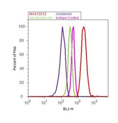
- Flow cytometry analysis of CD79a was done on Raji cells. Cells were fixed with 70% ethanol for 10 minutes, permeabilized with 0.25% Triton™ X-100 for 20 minutes, and blocked with 5% BSA for 30 minutes at room temperature. Cells were labeled with CD79a Mouse Monoclonal Antibody (Product # MA5-13212, red histogram) or with mouse isotype control (pink histogram) at 3-5 µg/million cells in 2.5% BSA. After incubation at room temperature for 2 hours, the cells were labeled with Alexa Fluor® 488 Rabbit Anti-Mouse Secondary Antibody (Product # A11059) at a dilution of 1:400 for 30 minutes at room temperature. The representative 10,000 cells were acquired and analyzed for each sample using an Attune® Acoustic Focusing Cytometer. The purple histogram represents unstained control cells and the green histogram represents no-primary-antibody control..
 Explore
 Validate
 Learn
Learn Western blot