Explore
 Validate
 Learn
Learn Western blot
Antibody data
- Antibody Data
- Antigen structure
- References [0]
- Comments [0]
- Validations
- Western blot [2]
- Immunohistochemistry [1]
Submit
Validation data
Reference
Comment
Report error
- Product number
- MAB8027 - Provider product page

- Provider
- R&D Systems
- Product name
- Anti-Human/Mouse/Rat Glutamate Dehydrogenase 2/GLUD2 Monoclonal Antibody (Clone 848209)
- Antibody type
- Monoclonal
- Antigen
- E. coli-derived recombinant human Glutamate Dehydrogenase 2/GLUD2, Ser54-Thr558
- Description
- Protein A or G purified from hybridoma culture supernatant
- Reactivity
- Human, Mouse, Rat
- Host
- Mouse
- Antigen sequence
P49448- Isotype
- IgG
- Vial size
- 100 µg
No comments: Submit comment
Supportive validation
- Submitted by
- R&D Systems (provider)
- Main image
- Experimental details
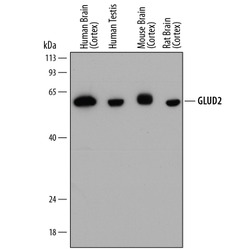
- Detection of Human, Mouse, and Rat GLUD2 by Western Blot. Western blot shows lysates of human brain (cortex) tissue, human testis tissue, mouse brain (cortex) tissue, and rat brain (cortex) tissue. PVDF membrane was probed with 0.2 µg/mL of Mouse Anti-Human/Mouse/Rat GLUD1/GLUD2 Monoclonal Antibody (Catalog # MAB8027) followed by HRP-conjugated Anti-Mouse IgG Secondary Antibody (Catalog # HAF018). A specific band was detected for GLUD2 at approximately 58 kDa (as indicated). This experiment was conducted under reducing conditions and using Immunoblot Buffer Group 1.

- Submitted by
- R&D Systems (provider)
- Main image

- Experimental details
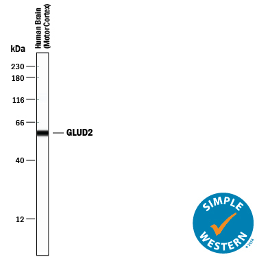
- Detection of Human GLUD2 by Simple WesternTM. Simple Western lane view shows lysates of human brain (motor cortex) tissue, loaded at 0.5 mg/mL. A specific band was detected for GLUD2 at approximately 58 kDa (as indicated) using 2 µg/mL of Mouse Anti-Human/Mouse/Rat GLUD1/GLUD2 Monoclonal Antibody (Catalog # MAB8027). This experiment was conducted under reducing conditions and using the 12-230 kDa separation system.
Supportive validation
- Submitted by
- R&D Systems (provider)
- Main image
- Experimental details
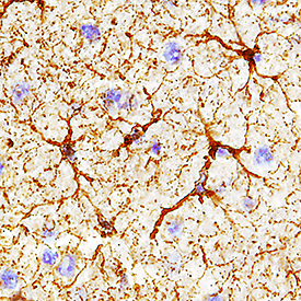
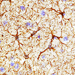
- GLUD1/GLUD2 in Rat Brain. GLUD1/GLUD2 was detected in perfusion fixed frozen sections of rat brain using Mouse Anti-Human/Mouse/Rat GLUD1/GLUD2 Monoclonal Antibody (Catalog # MAB8027) at 0.2 µg/mL for 1 hour at room temperature followed by incubation with the Anti-Mouse IgG VisUCyte™ HRP Polymer Antibody (Catalog # VC001). Tissue was stained using DAB (brown) and counterstained with hematoxylin (blue). Specific staining was localized to astrocytes. View our protocol for IHC Staining with VisUCyte HRP Polymer Detection Reagents.