 Explore
 Validate
 Learn
Learn Western blot
 Immunoprecipitation
Antibody data
- Antibody Data
- Antigen structure
- References [0]
- Comments [0]
- Validations
- Western blot [2]
- Immunohistochemistry [1]
- Other assay [1]
Submit
Validation data
Reference
Comment
Report error
- Product number
- AGC-010-200UL - Provider product page

- Provider
- Invitrogen Antibodies
- Product name
- GluR3 (GluA3) (extracellular) Polyclonal Antibody
- Antibody type
- Polyclonal
- Antigen
- Other
- Reactivity
- Human, Mouse, Rat
- Host
- Rabbit
- Isotype
- IgG
- Vial size
- 200 µL
- Concentration
- 0.6 mg/mL
- Storage
- -20° C, Avoid Freeze/Thaw Cycles
No comments: Submit comment
Supportive validation
- Submitted by
- Invitrogen Antibodies (provider)
- Main image
- Experimental details
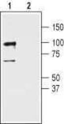
- Western blot analysis of rat cerebellum lysates: - 1. Anti-GluR3 (GluA3) (extracellular) Antibody (#AGC-010), (1:400). 2. Anti-GluR3 (GluA3) (extracellular) Antibody , preincubated with GluR3/GluA3 (extracellular) Blocking Peptide (#BLP-GC010).
- Submitted by
- Invitrogen Antibodies (provider)
- Main image

- Experimental details
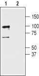
- Western blot analysis of rat cerebellum lysates: - 1. Anti-GluR3 (GluA3) (extracellular) Antibody (#AGC-010), (1:400). 2. Anti-GluR3 (GluA3) (extracellular) Antibody , preincubated with GluR3/GluA3 (extracellular) Blocking Peptide (#BLP-GC010).
Supportive validation
- Submitted by
- Invitrogen Antibodies (provider)
- Main image
- Experimental details
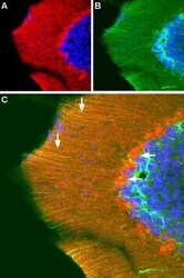
- Expression of GRIA3 (GluR3) in mouse cerebellum - Immunohistochemical stainingof frozen perfusion-fixed free floating sections of mouse cerebellum using Anti-GluR3 (GluA3) (extracellular) Antibody (#AGC-010). A. Distribution of GRIA3 (red). B. Distribution of glial fibrillary acidic protein (green). C. Merge of the two images indicates that GRIA3 is localized to Bergmann glia (vertical arrow) and to Purkinje cell soma (horizontal arrow). DAPI is used as the counterstain (blue).

Supportive validation
- Submitted by
- Invitrogen Antibodies (provider)
- Main image
- Experimental details
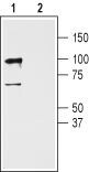
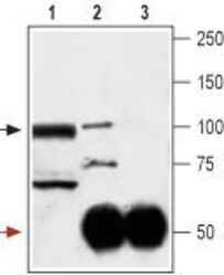
- Immunoprecipitation of rat cerebellum lysates: - 1. Cerebellum lysates. 2. Cerebellum lysates + Anti-GluR3 (GluA3) (extracellular) Antibody (#AGC-010) + protein A beads.3. Cerebellum lysates + pre-immune rabbit serum + protein A beads. Black arrow indicates GluR3 while the red arrow shows the IgG heavy chain. Immunoblot was performed with Anti-GluR3 (GluA3) (extracellular) Antibody (#AGC-010).
