Explore
 Validate
 Learn
LearnHPA030046
antibody from Atlas Antibodies
Targeting: USP48
FLJ11328, FLJ20103, FLJ23054, FLJ23277, MGC14879, USP31
 Western blot
Antibody data
- Antibody Data
- Antigen structure
- References [0]
- Comments [0]
- Validations
- Western blot [2]
- Immunocytochemistry [1]
- Immunohistochemistry [8]
Submit
Validation data
Reference
Comment
Report error
- Product number
- HPA030046 - Provider product page

- Provider
- Atlas Antibodies
- Proper citation
- Atlas Antibodies Cat#HPA030046, RRID:AB_10603325
- Product name
- Anti-USP48
- Antibody type
- Polyclonal
- Reactivity
- Human, Mouse, Rat
- Host
- Rabbit
- Conjugate
- Unconjugated
- Antigen sequence
QEKPNTTVQVPAFLQELVDRDNSKFEEWCIEMAEM
RKQSVDKGKAKHEEVKELYQRLPAGAEPYEFVSLE
WLQKWLDESTPTKPIDNHACLCSHDKLHPDKISIM
KRISEYAADIFYSRYGGGPRLTVKALCKECVV- Isotype
- IgG
- Vial size
- 100 µl
- Storage
- Store at +4°C for short term storage. Long time storage is recommended at -20°C.
No comments: Submit comment
Supportive validation
- Submitted by
- Atlas Antibodies (provider)
- Main image
- Experimental details
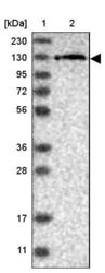
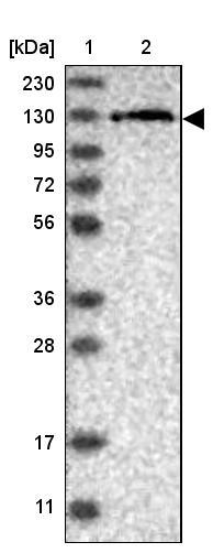
- Lane 1: Marker [kDa] 230, 130, 95, 72, 56, 36, 28, 17, 11Lane 2: Human cell line RT-4
- Submitted by
- Atlas Antibodies (provider)
- Main image
- Experimental details
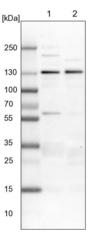
- Lane 1: NIH-3T3 cell lysate (Mouse embryonic fibroblast cells)Lane 2: NBT-II cell lysate (Rat Wistar bladder tumour cells)

Supportive validation
- Submitted by
- Atlas Antibodies (provider)
- Main image

- Experimental details
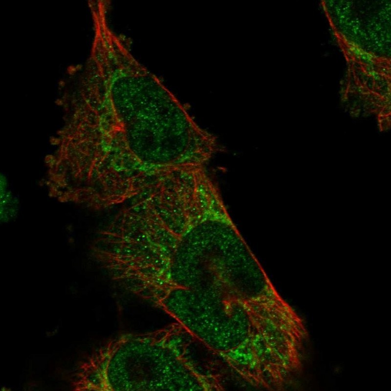
- Immunofluorescent staining of human cell line U-251 MG shows localization to nucleoplasm & mitochondria.
- Sample type
- HUMAN
Supportive validation
- Submitted by
- Atlas Antibodies (provider)
- Main image

- Experimental details
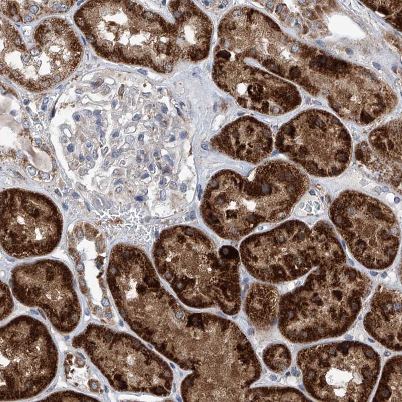
- Immunohistochemical staining of human kidney shows strong cytoplasmic positivity, with a granular pattern in cells in tubules.
- Sample type
- HUMAN
- Submitted by
- Atlas Antibodies (provider)
- Main image

- Experimental details
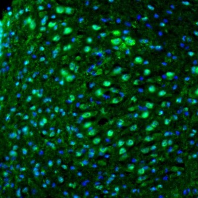
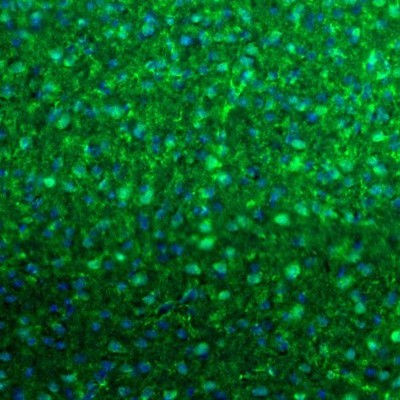
- Immunofluorescence staining of mouse pons shows immunoreactivity in locus coeruleus neurons.
- Sample type
- MOUSE
- Submitted by
- Atlas Antibodies (provider)
- Main image

- Experimental details
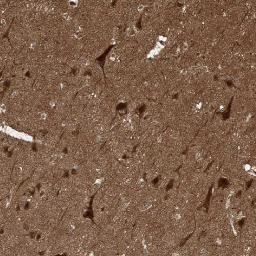
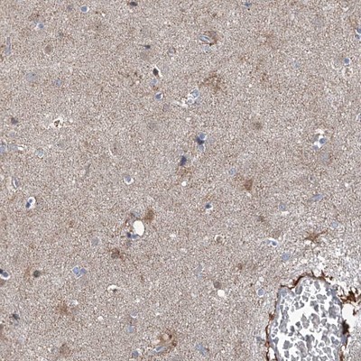
- Immunohistochemical staining of human lateral ventricle shows positivity in a subset of neurons as well as neuropil with a accentuation around blood vessels.
- Sample type
- HUMAN
- Submitted by
- Atlas Antibodies (provider)
- Main image

- Experimental details
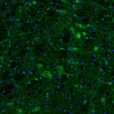
- Immunofluorescence staining of mouse visual cortex shows immunoreactivity in neurons and endothelial cells.
- Sample type
- MOUSE
- Submitted by
- Atlas Antibodies (provider)
- Main image

- Experimental details
- Immunofluorescence staining of mouse medulla shows neuronal staining in the facial nucleus.
- Sample type
- MOUSE
- Submitted by
- Atlas Antibodies (provider)
- Main image

- Experimental details
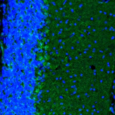
- Immunofluorescence staining of mouse cerebellum shows positivity in Purkinje cells.
- Sample type
- MOUSE
- Submitted by
- Atlas Antibodies (provider)
- Main image

- Experimental details
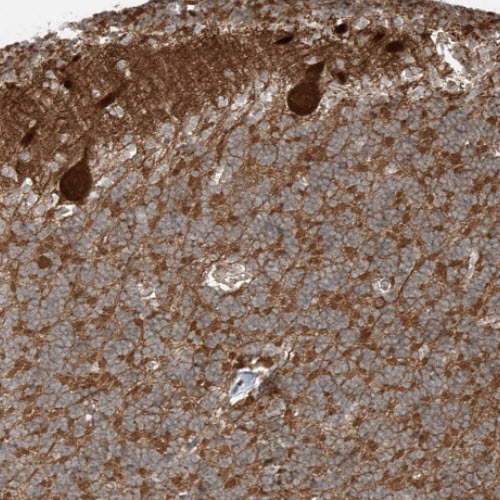
- Immunohistochemical staining of human cerebellum shows strong cytoplasmic positivity in Purkinje cells.
- Sample type
- HUMAN
- Submitted by
- Atlas Antibodies (provider)
- Main image

- Experimental details
- Immunohistochemical staining of human cerebral cortex shows cytoplasmic immunoreactivity in neurons, and positivity in neuropil.
- Sample type
- HUMAN