Explore
 Validate
 Learn
Learn Western blot
Antibody data
- Antibody Data
- Antigen structure
- References [0]
- Comments [0]
- Validations
- Western blot [1]
- Immunocytochemistry [1]
- Immunohistochemistry [2]
Submit
Validation data
Reference
Comment
Report error
- Product number
- 702618 - Provider product page

- Provider
- Invitrogen Antibodies
- Product name
- MCHR1 Recombinant Rabbit Monoclonal Antibody (12H4L9)
- Antibody type
- Monoclonal
- Antigen
- Synthetic peptide
- Description
- This antibody is predicted to react with Monkey, Mouse, Rabbit.
- Antibody clone number
- 12H4L9
- Concentration
- 0.5 mg/mL
No comments: Submit comment
Supportive validation
- Submitted by
- Invitrogen Antibodies (provider)
- Main image

- Experimental details
- Western blot analysis was performed on HEK-293 cells stably transfected with human MCHR1 (Lane 2) and mock (Lane 1). Receptors were eluted from the beads using SDS sample buffer for 20 min at 45 °C. Extracts were separated on 7.5% SDS-polyacrylamide gels and blotted onto PVDF membranes. The blot was probed with Anti-MCHR1 Recombinant Rabbit Monoclonal Antibody (Product # 702618, 1 µg/mL, 1:500 dilution). An ~55-70 kDa band corresponding to MCHR1 was observed only in the transfected cell line.

Supportive validation
- Submitted by
- Invitrogen Antibodies (provider)
- Main image
- Experimental details
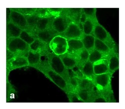
- For immunofluorescence analysis, HEK-293 cells were stably transfected with human MCHR1. After 24hr of transfection the cells were fixed and permeabilized for detection of MCHR1 using Anti-MCHR1 Recombinant Rabbit Monoclonal Antibody (Product # 702618, 1 µg/mL, 1:500 dilution) and labeled with Goat anti-Rabbit IgG (H+L) Superclonal™ Secondary Antibody, Alexa Fluor® 488 conjugate. Panel a) shows representative cells that were stained for detection of membrane localization of the MCHR1 on the transfected cells. The images were captured at 40X magnification.

Supportive validation
- Submitted by
- Invitrogen Antibodies (provider)
- Main image

- Experimental details
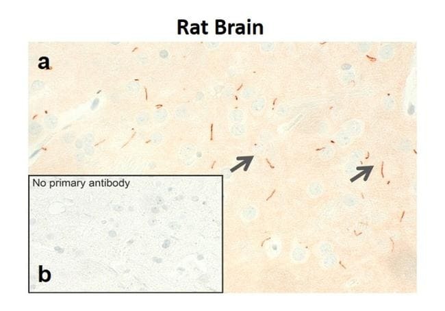
- Sections of Rat Brain was dewaxed, microwaved in citric acid and incubated with Anti-MCHR1 Recombinant Rabbit Monoclonal Antibody (Product # 702618, 1 µg/mL, 1:500 dilution). Sections were then sequentially treated with biotinylated Anti-rabbit IgG and AB solution. Sections were further developed in AEC and lightly counterstained with hematoxylin. Panel a) shows the specific localization of MCHR1 in the primary cilia on neuronal cells in Rat Brain. Panel b) shows the no primary antibody control.

- Submitted by
- Invitrogen Antibodies (provider)
- Main image

- Experimental details
- Sections of Rat Brain was dewaxed, microwaved in citric acid and incubated with Anti-MCHR1 Recombinant Rabbit Monoclonal Antibody (Product # 702618, 1 µg/mL, 1:500 dilution). Sections were then sequentially treated with biotinylated Anti-rabbit IgG and AB solution. Sections were further developed in AEC and lightly counterstained with hematoxylin. Panel a) shows the specific localization of MCHR1 in the primary cilia on neuronal cells in Rat Brain. Panel b) shows the no primary antibody control.