Explore
 Validate
 Learn
Learn Western blot
Antibody data
- Antibody Data
- Antigen structure
- References [0]
- Comments [0]
- Validations
- Western blot [2]
- Immunocytochemistry [1]
Submit
Validation data
Reference
Comment
Report error
- Product number
- 702394 - Provider product page

- Provider
- Invitrogen Antibodies
- Product name
- CDK5RAP2 Recombinant Rabbit Monoclonal Antibody (13H61L16)
- Antibody type
- Monoclonal
- Antigen
- Other
- Description
- This antibody is predicted to react with Monkey, Cat, Rat
- Antibody clone number
- 13H61L16
- Concentration
- 0.5 mg/mL
No comments: Submit comment
Supportive validation
- Submitted by
- Invitrogen Antibodies (provider)
- Main image
- Experimental details
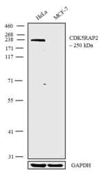
- Western blot analysis was performed on membrane extracts (30 µg lysate) of HeLa (Lane 1) and MCF-7 (Lane 2). The blots were probed with Anti-CDK5RAP2 Recombinant Rabbit Monoclonal Antibody (Product # 702394, 1-2 µg/mL). A 250 kDa band corresponding to CDK5RAP2 was observed as shown. The blots were detected by chemiluminescence using Goat anti-Rabbit IgG (H+L) Superclonal™ Secondary Antibody, HRP conjugate (Product # A27036, 0.4 µg/mL, 1:5000 dilution). Known quantity of protein samples were electrophoresed using Novex® NuPAGE® 4-12% Bis-Tris gel (Product # NP0321BOX), XCell SureLock™ Electrophoresis System (Product # EI0002) and Novex® Sharp Pre-Stained Protein Standard (Product # LC5800). Resolved proteins were then transferred onto a nitrocellulose membrane with wet transfer. The membrane was probed with the relevant primary and secondary Antibody following blocking with 5% skimmed milk. Chemiluminescent detection was performed using Pierce™ ECL Western blotting Substrate (Product # 32106).
- Submitted by
- Invitrogen Antibodies (provider)
- Main image

- Experimental details
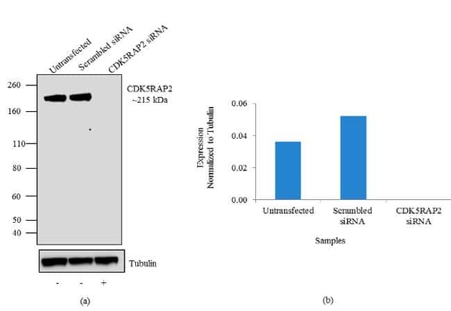
- Knockdown of CDK5RAP2 was achieved by transfecting HeLa cells with CDK5RAP2 specific siRNA (Silencer® select Cat # s31430 and s31431). Western blot analysis (Fig a) was performed using TEB extract from the HeLa knock down cells (lane 3), non-specific scrambled siRNA transfected cells (lane 2) and untransfected cells (lane 1). The blots were probed with Anti-CDK5RAP2 Recombinant Rabbit Monoclonal Antibody (Product # 702394, 1-3 µg/mL) and Goat anti-Rabbit IgG (H+L) Superclonal™ Secondary Antibody, HRP conjugate (Product # A27036, 0.4 µg/mL, 1:5000 dilution). Densitometric analysis of this Western blot is shown in histogram (Fig b). Loss of signal upon siRNA mediated knock down confirms that antibody is specific to CDK5RAP2.
Supportive validation
- Submitted by
- Invitrogen Antibodies (provider)
- Main image

- Experimental details
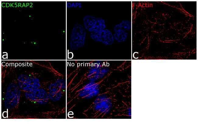
- For immunofluorescence analysis, HeLa cells were fixed and permeabilized for detection of endogenous CDK5RAP2 using Anti- CDK5RAP2 Recombinant Rabbit Monoclonal Antibody (Product # 702394, 2 µg/mL) and labeled with Goat anti-Rabbit IgG (H+L) Superclonal™ Secondary Antibody, Alexa Fluor® 488 conjugate (Product # A27034, 1:2000). Panel a) shows representative cells that were stained for detection and localization of CDK5RAP2 protein (green), Panel b) is stained for nuclei (blue) using SlowFade® Gold Antifade Mountant with DAPI (Product # S36938). Panel c) represents cytoskeletal F-actin staining using Rhodamine Phalloidin (Product # R415, 1:300). Panel d) is a composite image of panels a, b and c clearly demonstrating localization of CDK5RAP2 at the centrosomes. Panel e) represents control cells with no primary antibody to assess background. The images were captured at 60X magnification.