 Explore
 Validate
 Learn
Learn Western blot
Antibody data
- Antibody Data
- Antigen structure
- References [0]
- Comments [0]
- Validations
- Western blot [1]
- Immunohistochemistry [3]
Submit
Validation data
Reference
Comment
Report error
- Product number
- LS-C96481 - Provider product page

- Provider
- LSBio
- Product name
- EPHB2 / EPH Receptor B2 Antibody (clone 48CT12.6.4) LS-C96481
- Antibody type
- Monoclonal
- Description
- Protein G purified
- Reactivity
- Human
- Host
- Mouse
- Isotype
- IgG
- Antibody clone number
- 48CT12.6.4
- Storage
- Maintain refrigerated at 2°C to 8°C for up to 6 months. For long term storage store at -20°C.
No comments: Submit comment
Enhanced validation
- Submitted by
- LSBio (provider)
- Enhanced method
- Genetic validation
- Main image

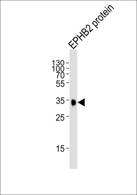
- Experimental details
- Western blot of lysate from EPHB2 protein, using EphB2 Antibody. Antibody was diluted at 1:1000. A goat anti-mouse IgG H&L (HRP) at 1:3000 dilution was used as the secondary antibody. Lysate at 35ug.
Enhanced validation
- Submitted by
- LSBio (provider)
- Enhanced method
- Genetic validation
- Main image

- Experimental details
- Formalin-fixed and paraffin-embedded human lung carcinoma tissue reacted with EPHB2 Monoclonal Antibody , which was peroxidase-conjugated to the secondary antibody, followed by DAB staining. This data demonstrates the use of this antibody for immunohistochemistry; clinical relevance has not been evaluated.

- Submitted by
- LSBio (provider)
- Enhanced method
- Genetic validation
- Main image
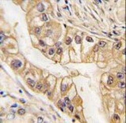
- Experimental details
- Formalin-fixed and paraffin-embedded human lung carcinoma tissue reacted with EPHB2 Monoclonal Antibody , which was peroxidase-conjugated to the secondary antibody, followed by DAB staining. This data demonstrates the use of this antibody for immunohistochemistry; clinical relevance has not been evaluated.
- Submitted by
- LSBio (provider)
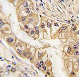
- Main image

- Experimental details
- Formalin-fixed and paraffin-embedded human lung carcinoma tissue reacted with EPHB2 Monoclonal Antibody , which was peroxidase-conjugated to the secondary antibody, followed by DAB staining. This data demonstrates the use of this antibody for immunohistochemistry; clinical relevance has not been evaluated.
