Explore
 Validate
 Learn
Learn Immunocytochemistry
Antibody data
- Antibody Data
- Antigen structure
- References [1]
- Comments [0]
- Validations
- Immunocytochemistry [1]
- Flow cytometry [1]
Submit
Validation data
Reference
Comment
Report error
- Product number
- MAB467 - Provider product page

- Provider
- R&D Systems
- Product name
- Human/Mouse EphB2 Antibody
- Antibody type
- Monoclonal
- Description
- Protein A or G purified from hybridoma culture supernatant. Detects human and mouse EphB2 in direct ELISAs.
- Reactivity
- Human, Mouse
- Host
- Rat
- Conjugate
- Unconjugated
- Antigen sequence
P54763- Isotype
- IgG
- Antibody clone number
- 512012
- Vial size
- 100 ug
- Concentration
- LYOPH
- Storage
- Use a manual defrost freezer and avoid repeated freeze-thaw cycles. 12 months from date of receipt, -20 to -70 °C as supplied. 1 month, 2 to 8 °C under sterile conditions after reconstitution. 6 months, -20 to -70 °C under sterile conditions after reconstitution.
Submitted references Dysregulation of EGFR Pathway in EphA2 Cell Subpopulation Significantly Associates with Poor Prognosis in Colorectal Cancer.
De Robertis M, Loiacono L, Fusilli C, Poeta ML, Mazza T, Sanchez M, Marchionni L, Signori E, Lamorte G, Vescovi AL, Garcia-Foncillas J, Fazio VM
Clinical cancer research : an official journal of the American Association for Cancer Research 2017 Jan 1;23(1):159-170
Clinical cancer research : an official journal of the American Association for Cancer Research 2017 Jan 1;23(1):159-170
No comments: Submit comment
Supportive validation
- Submitted by
- R&D Systems (provider)
- Main image

- Experimental details
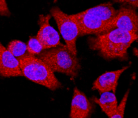
- EphB2 in MDA-MB-231 Human Cell Line. EphB2 was detected in immersion fixed MDA-MB-231 human breast cancer cell line using Rat Anti-Human/Mouse EphB2 Monoclonal Antibody (Catalog # MAB467) at 10 µg/mL for 3 hours at room temperature. Cells were stained using the NorthernLights™ 557-conjugated Anti-Rat IgG Secondary Antibody (red; Catalog # NL013) and counterstained with DAPI (blue). Specific staining was localized to cytoplasm. View our protocol for Fluorescent ICC Staining of Cells on Coverslips.
Supportive validation
- Submitted by
- R&D Systems (provider)
- Main image
- Experimental details
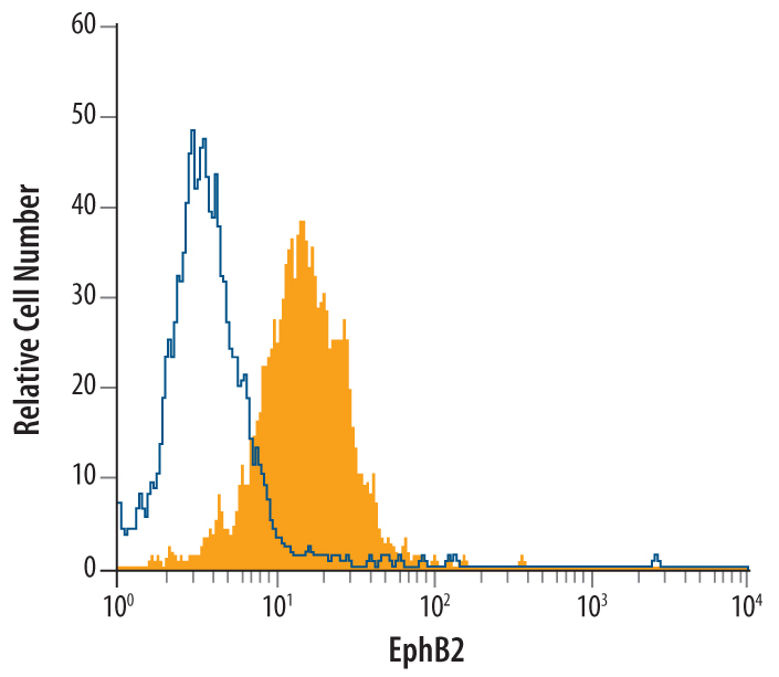
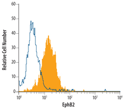
- Detection of EphB2 in MDA-MB-231 Human Cell Line by Flow Cytometry. MDA-MB-231 human breast cancer cell line was stained with Rat Anti-Mouse EphB2 Monoclonal Antibody (Catalog # MAB467, filled histogram) or isotype control antibody (Catalog # MAB006, open histogram), followed by Phycoerythrin-conjugated Anti-Rat IgG F(ab')2 Secondary Antibody (Catalog # F0105B).