 Explore
 Validate
 Learn
Learn Western blot
 Immunohistochemistry
Antibody data
- Antibody Data
- Antigen structure
- References [0]
- Comments [0]
- Validations
- Western blot [2]
- Immunocytochemistry [1]
- Immunohistochemistry [10]
Submit
Validation data
Reference
Comment
Report error
- Product number
- HPA031219 - Provider product page

- Provider
- Atlas Antibodies
- Proper citation
- Atlas Antibodies Cat#HPA031219, RRID:AB_10601989
- Product name
- Anti-BYSL
- Antibody type
- Polyclonal
- Reactivity
- Human
- Host
- Rabbit
- Conjugate
- Unconjugated
- Antigen sequence
QQQEELEAEHGTGDKPAAPRERTTRLGPRMPQDGS
DDEDEEWPTLEKAATMTAAGHHAEVVVDPEDERAI
EMFMNKNPPAR- Isotype
- IgG
- Vial size
- 100 µl
- Storage
- Store at +4°C for short term storage. Long time storage is recommended at -20°C.
No comments: Submit comment
Supportive validation
Supportive validation
- Submitted by
- Atlas Antibodies (provider)
- Enhanced method
- Independent antibody validation
- Main image

- Experimental details
- Western blot analysis using Anti-BYSL antibody HPA031219 (A) shows similar pattern to independent antibody HPA031217 (B).

Supportive validation
- Submitted by
- Atlas Antibodies (provider)
- Main image

- Experimental details
- Lane 1: Marker [kDa] 230, 130, 95, 72, 56, 36, 28, 17, 11Lane 2: Human cell line RT-4

Supportive validation
- Submitted by
- Atlas Antibodies (provider)
- Main image

- Experimental details
- Immunofluorescent staining of human cell line U-2 OS shows localization to nucleus, nucleoli & vesicles.
- Sample type
- HUMAN

Enhanced validation
Supportive validation
- Submitted by
- Atlas Antibodies (provider)
- Enhanced method
- Independent antibody validation
- Main image

- Experimental details
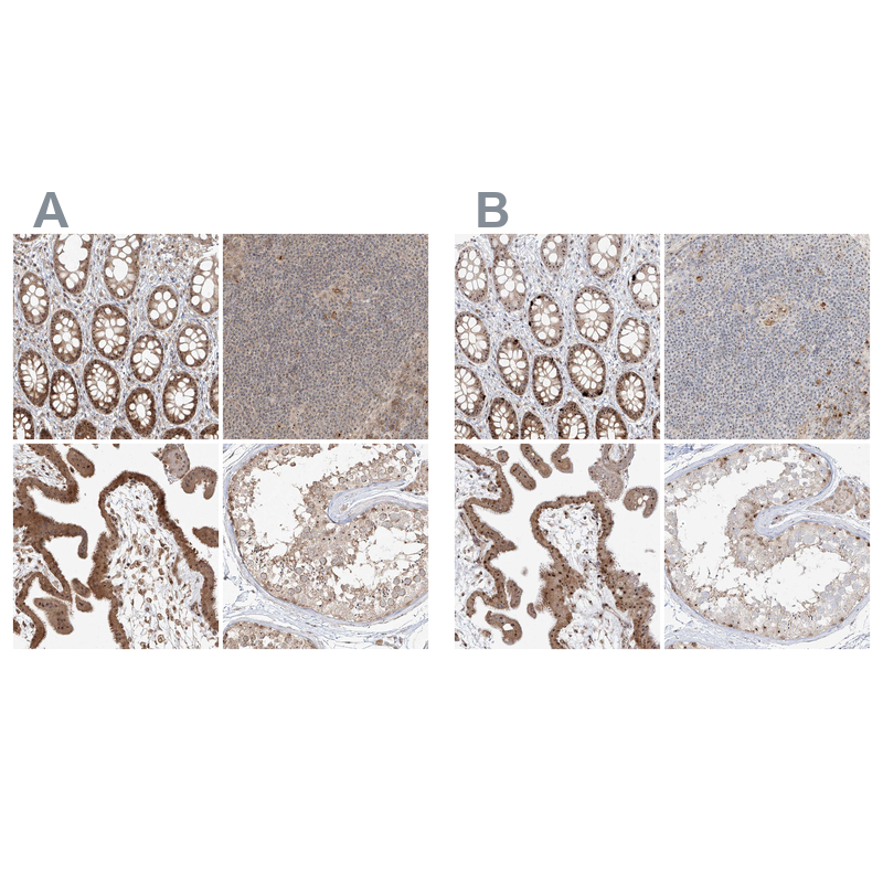
- Immunohistochemical staining of human gastrointestinal, lymphoid tissues, placenta and testis using Anti-BYSL antibody HPA031219 (A) shows similar protein distribution across tissues to independent antibody HPA031217 (B).
Supportive validation
- Submitted by
- Atlas Antibodies (provider)
- Main image
- Experimental details
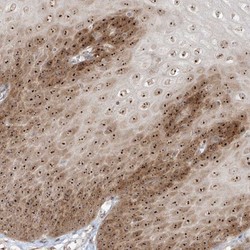
- Immunohistochemical staining of human esophagus shows strong nucleolar and cytoplasmic positivity in squamous epithelial cells.

- Submitted by
- Atlas Antibodies (provider)
- Main image
- Experimental details
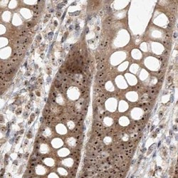
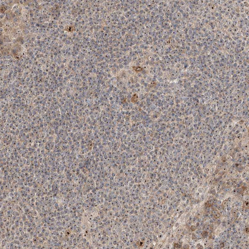
- Immunohistochemical staining of human colon using Anti-BYSL antibody HPA031219.
- Sample type
- HUMAN

- Submitted by
- Atlas Antibodies (provider)
- Main image

- Experimental details
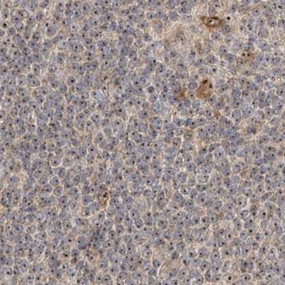
- Immunohistochemical staining of human lymph node using Anti-BYSL antibody HPA031219.
- Sample type
- HUMAN
- Submitted by
- Atlas Antibodies (provider)
- Main image

- Experimental details
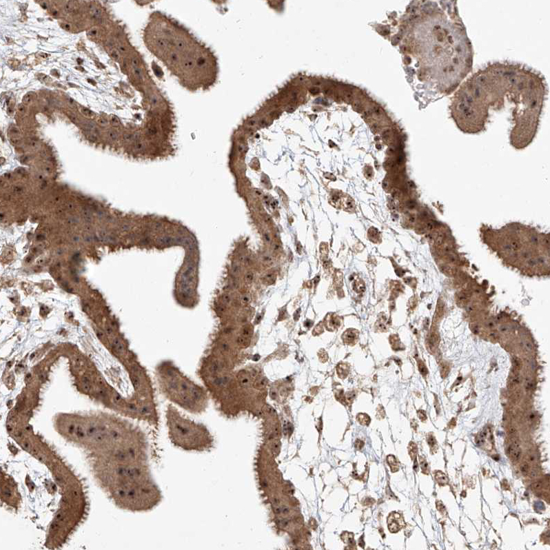
- Immunohistochemical staining of human placenta using Anti-BYSL antibody HPA031219.
- Sample type
- HUMAN

- Submitted by
- Atlas Antibodies (provider)
- Main image

- Experimental details
- Immunohistochemical staining of human testis using Anti-BYSL antibody HPA031219.
- Sample type
- HUMAN

- Submitted by
- Atlas Antibodies (provider)
- Main image

- Experimental details
- Immunohistochemical staining of human testis shows moderate positivity in nucleoli in cells in seminiferous ducts.
- Sample type
- HUMAN

- Submitted by
- Atlas Antibodies (provider)
- Main image

- Experimental details
- Immunohistochemical staining of human placenta shows strong positivity in nucleoli in trophoblastic cells.
- Sample type
- HUMAN
- Submitted by
- Atlas Antibodies (provider)
- Main image

- Experimental details
- Immunohistochemical staining of human lymph node shows strong positivity in nucleoli in lymphoid cells.
- Sample type
- HUMAN
- Submitted by
- Atlas Antibodies (provider)
- Main image

- Experimental details
- Immunohistochemical staining of human rectum shows strong positivity in nucleoli in glandular cells.
- Sample type
- HUMAN
