 Explore
 Validate
 Learn
LearnPA3-16805
antibody from Invitrogen Antibodies
Targeting: SCARB2
CD36L2, HLGP85, LIMP-2, LIMPII, SR-BII
 Western blot Immunocytochemistry
 Immunoprecipitation Immunohistochemistry Flow cytometry Blocking/Neutralizing
Antibody data
- Antibody Data
- Antigen structure
- References [1]
- Comments [0]
- Validations
- Western blot [1]
- Immunocytochemistry [1]
- Immunohistochemistry [2]
- Flow cytometry [1]
Submit
Validation data
Reference
Comment
Report error
- Product number
- PA3-16805 - Provider product page

- Provider
- Invitrogen Antibodies
- Product name
- SR-BI/SR-BII Polyclonal Antibody
- Antibody type
- Polyclonal
- Antigen
- Other
- Reactivity
- Human, Mouse, Rat
- Host
- Rabbit
- Isotype
- IgG
- Vial size
- 100 µL
- Concentration
- Conc. Not Determined
- Storage
- -20° C, Avoid Freeze/Thaw Cycles
Submitted references Zwitterionic hydrogels implanted in mice resist the foreign-body reaction.
Zhang L, Cao Z, Bai T, Carr L, Ella-Menye JR, Irvin C, Ratner BD, Jiang S
Nature biotechnology 2013 Jun;31(6):553-6
Nature biotechnology 2013 Jun;31(6):553-6
No comments: Submit comment
Supportive validation
- Submitted by
- Invitrogen Antibodies (provider)
- Main image

- Experimental details
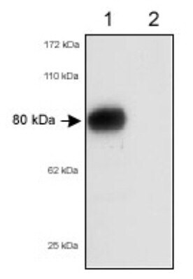
- Western blot analysis of SR-BI/SR-BII in 80 µg total mouse liver lysates. Samples were incubated in SR-BI/SR-BII polyclonal antibody (Product # PA3-16805). Lane 1: wild-type mice; Lane 2: SR-BI deficient mice.
Supportive validation
- Submitted by
- Invitrogen Antibodies (provider)
- Main image

- Experimental details
- Immunocytochemistry analysis of SR-BI/SR-BII in HeLa cells. Samples were incubated in SR-BI/SR-BII polyclonal antibody (Product # PA3-16805) followed by DyLight 488 (green). Nuclei and alpha-tubulin were counterstained with DAPI (blue) and DyLight 550 (red).

Supportive validation
- Submitted by
- Invitrogen Antibodies (provider)
- Main image

- Experimental details
- Immunohistochemical analysis of SR-BI/SR-BII in formalin-fixed paraffin-embedded tissue section of normal human liver. Samples were incubated in SR-BI/SR-BII polyclonal antibody (Product # PA3-16805) using a dilution of 5 µg/mL. Specific and expected granular membrane-cytoplasmic staining was observed in the hepatocytes. [40X Magnification].

- Submitted by
- Invitrogen Antibodies (provider)
- Main image

- Experimental details
- SR-BI/II staining in mouse subcutaneous tissue post-injury. Tissues were fixed in Zinc-Tris buffer overnight, blocked with 2.5% rabbit serum, and stained with Scavenger Receptor B1/2 Polyclonal Antibody (Product # PA3-16805) at 1:1000 dilution followed by a biotinylated-goat anti-rabbit IgG secondary antibody, avidin-biotin reagent and DAB substrate. Data courtesy of the Innovators Program

Supportive validation
- Submitted by
- Invitrogen Antibodies (provider)
- Main image
- Experimental details
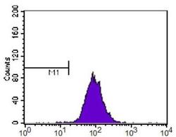
- Flow cytometry of SR-BI/SR-BII in NIH-3T3 cells. Samples were incubated in SR-BI/SR-BII polyclonal antibody (Product # PA3-16805) using a dilution of 1:400 followed by a Alexa Fluor 488 secondary (shown in purple). M1 is defined by unstained cells.
