 Explore
 Validate
 Learn
Learn Western blot
Antibody data
- Antibody Data
- Antigen structure
- References [8]
- Comments [0]
- Validations
- Western blot [2]
- Immunocytochemistry [1]
- Immunohistochemistry [1]
- Flow cytometry [1]
- Other assay [2]
Submit
Validation data
Reference
Comment
Report error
- Product number
- 701457 - Provider product page

- Provider
- Invitrogen Antibodies
- Product name
- Alpha-Smooth Muscle Actin Recombinant Rabbit Monoclonal Antibody (17H19L35)
- Antibody type
- Monoclonal
- Antigen
- Synthetic peptide
- Description
- This antibody is predicted to react with bovine, canine, rabbit, rat and porcine based on sequence homology.
- Antibody clone number
- 17H19L35
- Concentration
- 0.5 mg/mL
Submitted references Multiple benign fibrous histiocytomas of the mandible: A case report and review of the literature.
Single Cell Analysis of Cultivated Fibroblasts from Chronic Pancreatitis and Pancreatic Cancer Patients.
L-carnitine suppresses transient receptor potential vanilloid type 1 activity and myofibroblast transdifferentiation in human corneal keratocytes.
Purine nucleoside phosphorylase inhibition ameliorates age-associated lower urinary tract dysfunctions.
TFAP2C facilitates somatic cell reprogramming by inhibiting c-Myc-dependent apoptosis and promoting mesenchymal-to-epithelial transition.
Eugenol, a potential schistosomicidal agent with anti-inflammatory and antifibrotic effects against Schistosoma mansoni, induced liver pathology.
Severe pulmonary hypertension in aging female apolipoprotein E-deficient mice is rescued by estrogen replacement therapy.
CD98 regulates vascular smooth muscle cell proliferation in atherosclerosis.
Wang Y, Huang Y, Cai WX, Tao Q
Experimental and therapeutic medicine 2022 Sep;24(3):593
Experimental and therapeutic medicine 2022 Sep;24(3):593
Single Cell Analysis of Cultivated Fibroblasts from Chronic Pancreatitis and Pancreatic Cancer Patients.
Sunami Y, Chen Y, Trojanowicz B, Sommerer M, Hämmerle M, Eils R, Kleeff J
Cells 2022 Aug 19;11(16)
Cells 2022 Aug 19;11(16)
L-carnitine suppresses transient receptor potential vanilloid type 1 activity and myofibroblast transdifferentiation in human corneal keratocytes.
Turan E, Valtink M, Reinach PS, Skupin A, Luo H, Brockmann T, Ba Salem MHO, Pleyer U, Mergler S
Laboratory investigation; a journal of technical methods and pathology 2021 Jun;101(6):680-689
Laboratory investigation; a journal of technical methods and pathology 2021 Jun;101(6):680-689
Purine nucleoside phosphorylase inhibition ameliorates age-associated lower urinary tract dysfunctions.
Birder LA, Wolf-Johnston A, Wein AJ, Cheng F, Grove-Sullivan M, Kanai AJ, Watson AM, Stoltz D, Watkins SC, Robertson AM, Newman D, Dmochowski RR, Jackson EK
JCI insight 2020 Oct 15;5(20)
JCI insight 2020 Oct 15;5(20)
TFAP2C facilitates somatic cell reprogramming by inhibiting c-Myc-dependent apoptosis and promoting mesenchymal-to-epithelial transition.
Wang Y, Chen S, Jiang Q, Deng J, Cheng F, Lin Y, Cheng L, Ye Y, Chen X, Yao Y, Zhang X, Shi G, Dai L, Su X, Peng Y, Deng H
Cell death & disease 2020 Jun 25;11(6):482
Cell death & disease 2020 Jun 25;11(6):482
Eugenol, a potential schistosomicidal agent with anti-inflammatory and antifibrotic effects against Schistosoma mansoni, induced liver pathology.
El-Kady AM, Ahmad AA, Hassan TM, El-Deek HEM, Fouad SS, Althagfan SS
Infection and drug resistance 2019;12:709-719
Infection and drug resistance 2019;12:709-719
Severe pulmonary hypertension in aging female apolipoprotein E-deficient mice is rescued by estrogen replacement therapy.
Umar S, Partow-Navid R, Ruffenach G, Iorga A, Moazeni S, Eghbali M
Biology of sex differences 2017;8:9
Biology of sex differences 2017;8:9
CD98 regulates vascular smooth muscle cell proliferation in atherosclerosis.
Baumer Y, McCurdy S, Alcala M, Mehta N, Lee BH, Ginsberg MH, Boisvert WA
Atherosclerosis 2017 Jan;256:105-114
Atherosclerosis 2017 Jan;256:105-114
No comments: Submit comment
Supportive validation
- Submitted by
- Invitrogen Antibodies (provider)
- Main image

- Experimental details
- Western blot analysis of Smooth Muscle Actin was performed by loading 20 µg of Rat Heart (lane1) and Mouse Heart (lane2) tissue lysates using Novex®NuPAGE®4-12 % Bis-Tris gel (Product # NP0321BOX), XCell SureLock Electrophoresis System (Product # EI0002), Novex® Sharp Pre-Stained Protein Standard (Product # LC5800), and iBlot® Dry Blotting System (Product # IB21001). Proteins were transferred to a nitrocellulose membrane and blocked with 5 % skim milk for 1 hour at room temperature. Smooth Muscle Actin was detected at ~40 kDa using Smooth Muscle Actin Recombinant Rabbit Monoclonal Antibody (Product # 701457) at 1 µg-3 µg/mL in 2.5 % skim milk at 4°C overnight on a rocking platform. Goat anti-Rabbit IgG-HRP Secondary Antibody (Product # G-21234) at 1:5000 dilution was used and chemiluminescent detection was performed using Pierce™ ECL Western blotting Substrate (Product # 32106).

- Submitted by
- Invitrogen Antibodies (provider)
- Main image

- Experimental details
- Western blot analysis of Smooth Muscle Actin in whole cell extracts from rat heart using a Smooth Muscle Actin recombinant rabbit monoclonal antibody (Product # 701457) at a dilution of 1 µg/mL. Detection was performed using an HRP-conjugated goat anti-rabbit secondary antibody followed by chemiluminescence (ECL). Results show a band at ~40kDa.

Supportive validation
- Submitted by
- Invitrogen Antibodies (provider)
- Main image

- Experimental details
- Immunofluorescent analysis of Smooth muscle actin was done on 70% confluent log phase MDCK cells. The cells were fixed with 4% paraformaldehyde for 15 minutes; permeabilized with 0.25% Triton X-100 for 10 minutes followed by blocking with 5% BSA for 1 hour at room temperature. The cells were incubated with Smooth muscle actin Recombinant Rabbit Monoclonal Antibody (Product # 701457) at 2 µg-4 µg in 1% BSA and incubated for 3 hours at room temperature and then labeled with Alexa Fluor® 488 Goat anti-Rabbit IgG Secondary Antibody (Product # A-11008) at a dilution of 1:400 for 30 minutes at room temperature (Panel a: green). Nuclei (Panel b: blue) were stained with SlowFade® Gold Antifade Mountant with DAPI (Product # S36938). F-actin (Panel c: red) was stained with Alexa Fluor® 594 Phalloidin (Product # A12381). Panel d is a merged image showing actin filament localization of Smooth muscle actin. Panel e shows no primary antibody control. The images were captured at 20X magnification.

Supportive validation
- Submitted by
- Invitrogen Antibodies (provider)
- Main image

- Experimental details
- Immunohistochemistry analysis of Smooth Muscle Actin showing staining in the cytoplasm of paraffin-embedded human uterus tissue (right) compared to a negative control without primary antibody (left). To expose target proteins, antigen retrieval was performed using 10 mM sodium citrate (pH 6.0), microwaved for 8-15 min. Following antigen retrieval, tissues were blocked in 3% H2O2-methanol for 15 min at room temperature, washed with ddH2O and PBS, and then probed with Smooth Muscle Actin monoclonal antibody (Product # 701457) diluted in 3% BSA-PBS at a dilution of 1:100 overnight at 4°C in a humidified chamber. Tissues were washed extensively in PBST and detection was performed using a HRP-conjugated secondary antibody followed by colorimetric detection using a DAB kit. Tissues were counterstained with hematoxylin and dehydrated with ethanol and xylene to prep for mounting.

Supportive validation
- Submitted by
- Invitrogen Antibodies (provider)
- Main image

- Experimental details
- Flow cytometry analysis of Smooth Muscle Actin was done on HeLa cells. Cells were fixed with 70% ethanol for 10 minutes, permeabilized with 0.25% Tritonª X-100 for 20 minutes, and blocked with 5% BSA for 1 hour at room temperature. Cells were labeled with ABfinityª Smooth Muscle Actin Recombinant Rabbit Monoclonal Antibody (701457, red histogram) or with rabbit isotype control (pink histogram) at 2 µg-4 µg/million cells in 2.5% BSA. After incubation at room temperature for 2-3 hours, the cells were labeled with Alexa Fluor¨ 488 Goat Anti-Rabbit Secondary Antibody (A11008) at a dilution of 1:400 for 30 minutes at room temperature. The representative 10,000 cells were acquired and analyzed for each sample using an Attune¨ Acoustic Focusing Cytometer. The purple histogram represents unstained control cells and the green histogram represents no-primary-antibody control.

Supportive validation
- Submitted by
- Invitrogen Antibodies (provider)
- Main image

- Experimental details
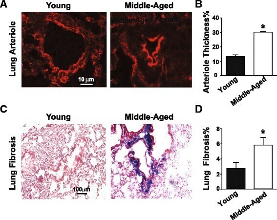
- Fig. 2 Development of pulmonary vascular remodeling and pulmonary fibrosis in middle-aged female ApoE-deficient mice. a Immunofluorescence images showing alpha-smooth muscle actin stained pulmonary arterioles in young and middle-aged female ApoE-deficient mice. b Quantification of arteriolar wall thickness for vessels less than 50 mum in young ( n = 3) and MA ( n = 3) ApoE-difiecent mice. * p < 0.05 vs. young female ( t test). Expressed as mean +- SEM. c Masson trichrome stained lung sections showing pulmonary fibrosis ( blue ) in young female and middle-aged female ApoE-deficient mice. d Quantification of pulmonary fibrosis in young ( n = 5) and MA ( n = 4) ApoE-deficient mice. * p < 0.05 vs. young female ( t test). Values are expressed as mean +- SEM
- Submitted by
- Invitrogen Antibodies (provider)
- Main image

- Experimental details
- Immunocytochemistry of alphaSMA and CXCL12 in disease-associated fibroblasts from pancreatic cancer and chronic pancreatitis patients. Cells from ( A ) patient1, ( B ) from patient2, and ( C ) from patient3 for alphaSMA staining, ( D ) cells from patient1, ( E ) from patient2, and ( F ) from patient3 for CXCL12 staining. Bar: 50 mum.
