Explore
 Validate
 Learn
Learn Western blot
 Immunoprecipitation
Antibody data
- Antibody Data
- Antigen structure
- References [0]
- Comments [0]
- Validations
- Western blot [4]
- Immunocytochemistry [2]
- Flow cytometry [1]
- Other assay [1]
Submit
Validation data
Reference
Comment
Report error
- Product number
- 16034-1-AP - Provider product page

- Provider
- Invitrogen Antibodies
- Product name
- PSMD7 Polyclonal Antibody
- Antibody type
- Polyclonal
- Antigen
- Other
- Description
- Immunogen sequence: MPELAVQKV VVHPLVLLSV VDHFNRIGKV GNQKRVVGVL LGSWQKKVLD VSNSFAVPFD EDDKDDSVWF LDHDYLENMY GMFKKVNARE RIVGWYHTGP KLHKNDIAIN ELMKRYCPNS VLVIIDVKPK DLGLPTEAYI SVEEVHDDGT PTSKTFEHVT SEIGAEEAEE VGVEHLLRDI KDTTVGTLSQ RITNQVHGLK GLNSKLLDIR SYLEKVATGK LPINHQIIYQ LQDVFNLLPD VSLQEFVKAF YLKTNDQMVV VYLASLIRSV VALHNLINNK IANRDAEKKE GQEKEESKKD RKEDKEKDKD KEKSDVKKEE (1-319 aa encoded by BC012606)
- Reactivity
- Human, Mouse, Rat
- Host
- Rabbit
- Isotype
- IgG
- Vial size
- 150 µL
- Concentration
- 0.13 mg/mL
- Storage
- -20°C
No comments: Submit comment
Supportive validation
- Submitted by
- Invitrogen Antibodies (provider)
- Main image

- Experimental details
- HeLa cells were subjected to SDS PAGE followed by western blot with 16034-1-AP (PSMD7 antibody) at dilution of 1:500 incubated at room temperature for 1.5 hours.

- Submitted by
- Invitrogen Antibodies (provider)
- Main image

- Experimental details
- Human liver tissue were subjected to SDS PAGE followed by western blot with 16034-1-AP (PSMD7 antibody) at dilution of 1:500 incubated at room temperature for 1.5 hours.

- Submitted by
- Invitrogen Antibodies (provider)
- Main image

- Experimental details
- Mouse liver tissue were subjected to SDS PAGE followed by western blot with 16034-1-AP ( PSMD7 Antibody) at dilution of 1:1200 incubated at room temperature for 1.5 hours.

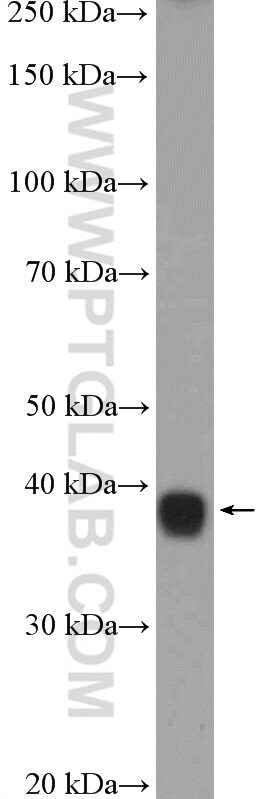
- Submitted by
- Invitrogen Antibodies (provider)
- Main image

- Experimental details
- Mouse liver tissue were subjected to SDS PAGE followed by western blot with 16034-1-AP (PSMD7 Antibody) at dilution of 1:600 incubated at room temperature for 1.5 hours.
Supportive validation
- Submitted by
- Invitrogen Antibodies (provider)
- Main image

- Experimental details
- Immunofluorescent analysis of HepG2 cells, using PSMD7 antibody 16034-1-AP at 1:25 dilution and Rhodamine-labeled goat anti-rabbit IGG (red).

- Submitted by
- Invitrogen Antibodies (provider)
- Main image
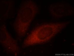
- Experimental details
- Immunofluorescent analysis of HepG2 cells, using PSMD7 antibody 16034-1-AP at 1:25 dilution and Rhodamine-labeled goat anti-rabbit IGG (red).

Supportive validation
- Submitted by
- Invitrogen Antibodies (provider)
- Main image
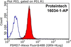
- Experimental details
- 1X10^6 HepG2 cells were stained with 0.2ug PSMD7 antibody (16034-1-AP, red) and control antibody (blue). Fixed with 90% MeOH blocked with 3% BSA (30 min). Alexa Fluor 488-conjugated AffiniPure Goat Anti-Rabbit IGG (H+L) with dilution 1:1500.

Supportive validation
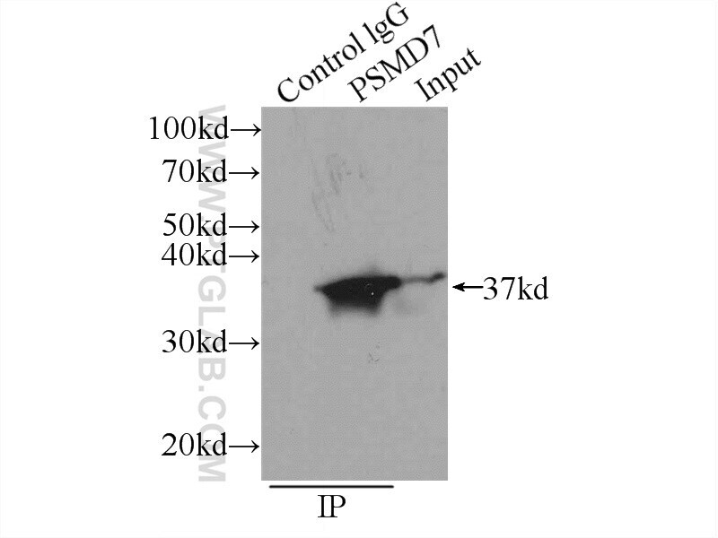
- Submitted by
- Invitrogen Antibodies (provider)
- Main image

- Experimental details
- IP result of anti-PSMD7 (IP:16034-1-AP, 3ug; Detection:16034-1-AP 1:500) with K-562 cells lysate 2400ug.