Explore
 Validate
 Learn
Learn Western blot
Antibody data
- Antibody Data
- Antigen structure
- References [1]
- Comments [0]
- Validations
- Western blot [2]
- Immunocytochemistry [2]
- Immunohistochemistry [4]
- Flow cytometry [1]
Submit
Validation data
Reference
Comment
Report error
- Product number
- TA500726 - Provider product page

- Provider
- OriGene
- Proper citation
- OriGene Cat#TA500726, RRID:AB_11127767
- Product name
- Anti-IFIT3 mouse monoclonal antibody, clone OTI1G1 (formerly 1G1)
- Antibody type
- Monoclonal
- Description
- Anti-IFIT3 mouse monoclonal antibody, clone OTI1G1 (formerly 1G1)
- Host
- Mouse
- Conjugate
- Unconjugated
- Epitope
- IFIT3
- Isotype
- IgG
- Antibody clone number
- OTI1G1
- Vial size
- 100 µl
- Concentration
- NULL
Submitted references Human IFIT3 Modulates IFIT1 RNA Binding Specificity and Protein Stability.
Johnson B, VanBlargan LA, Xu W, White JP, Shan C, Shi PY, Zhang R, Adhikari J, Gross ML, Leung DW, Diamond MS, Amarasinghe GK
Immunity 2018 Mar 20;48(3):487-499.e5
Immunity 2018 Mar 20;48(3):487-499.e5
No comments: Submit comment
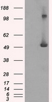
Supportive validation
- Submitted by
- OriGene (provider)
- Main image
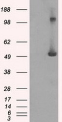
- Experimental details
- HEK293T cells were transfected with the pCMV6-ENTRY control (Left lane) or pCMV6-ENTRY IFIT3 (RC200610, Right lane) cDNA for 48 hrs and lysed. Equivalent amounts of cell lysates (5 ug per lane) were separated by SDS-PAGE and immunoblotted with anti-IFIT3.
- Validation comment
- WB
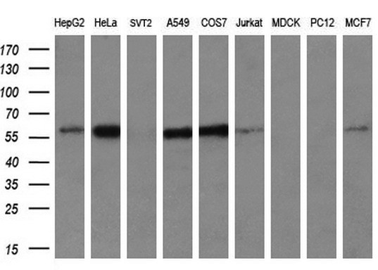
- Submitted by
- OriGene (provider)
- Main image

- Experimental details
- Western blot analysis of extracts (35ug) from 9 different cell lines by using anti-IFIT3 monoclonal antibody at 1:200 dilution. (HepG2: human; HeLa: human; SVT2: mouse; A549: human; COS7: monkey; Jurkat: human; MDCK: canine; PC12: rat; MCF7: human)
- Validation comment
- WB
Supportive validation
- Submitted by
- OriGene (provider)
- Main image

- Experimental details
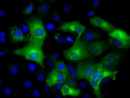
- Anti-IFIT3 mouse monoclonal antibody (TA500726) immunofluorescent staining of COS7 cells transiently transfected by pCMV6-ENTRY IFIT3(RC200610).
- Validation comment
- IF
- Submitted by
- OriGene (provider)
- Main image

- Experimental details
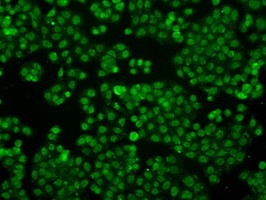
- Immunofluorescent staining of HT29 cells using anti-IFIT3 mouse monoclonal antibody (TA500726).
- Validation comment
- IF
Supportive validation
- Submitted by
- OriGene (provider)
- Main image
- Experimental details
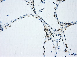
- Immunohistochemical staining of paraffin-embedded colon tissue within the normal limits using anti-IFIT3mouse monoclonal antibody. (Heat-induced epitope retrieval by 10mM citric buffer, pH6.0, 100C for 10min, TA500726, Dilution 1:50)
- Validation comment
- IHC
- Submitted by
- OriGene (provider)
- Main image
- Experimental details
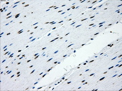
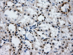
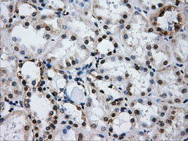
- Immunohistochemical staining of paraffin-embedded Kidney tissue within the normal limits using anti-IFIT3mouse monoclonal antibody. (Heat-induced epitope retrieval by 10mM citric buffer, pH6.0, 100C for 10min, TA500726, Dilution 1:50)
- Validation comment
- IHC
- Submitted by
- OriGene (provider)
- Main image
- Experimental details
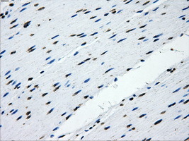
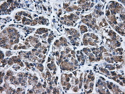
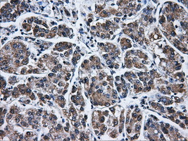
- Immunohistochemical staining of paraffin-embedded Carcinoma of liver tissue using anti-IFIT3mouse monoclonal antibody. (Heat-induced epitope retrieval by 10mM citric buffer, pH6.0, 100C for 10min, TA500726, Dilution 1:50)
- Validation comment
- IHC
- Submitted by
- OriGene (provider)
- Main image

- Experimental details
- Immunohistochemical staining of paraffin-embedded thyroid tissue within the normal limits using anti-IFIT3mouse monoclonal antibody. (Heat-induced epitope retrieval by 10mM citric buffer, pH6.0, 100C for 10min, TA500726, Dilution 1:50)
- Validation comment
- IHC
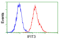
Supportive validation
- Submitted by
- OriGene (provider)
- Main image

- Experimental details
- Flow cytometric analysis of Jurkat cells, using anti-IFIT3 antibody(TA500726),(Red) compared to a nonspecific negative control antibody(TA50011)(Blue).
- Validation comment
- FC