Explore
 Validate
 Learn
Learn Western blot
Antibody data
- Antibody Data
- Antigen structure
- References [0]
- Comments [0]
- Validations
- Western blot [2]
- Immunocytochemistry [2]
- Immunohistochemistry [3]
Submit
Validation data
Reference
Comment
Report error
- Product number
- PA5-81131 - Provider product page

- Provider
- Invitrogen Antibodies
- Product name
- ZIC3 Polyclonal Antibody
- Antibody type
- Polyclonal
- Antigen
- Synthetic peptide
- Description
- This product is preservative free. It is recommended to add sodium azide to avoid contamination (final concentration 0.05%-0.1%).
- Concentration
- 5 mg/mL
No comments: Submit comment
Supportive validation
- Submitted by
- Invitrogen Antibodies (provider)
- Main image
- Experimental details
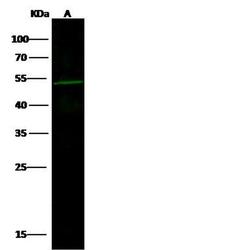
- Western blot analysis of ZIC3 in Lane A: Jurkat Whole Cell Lysate (30 µg). Samples were probed using a ZIC3 Polyclonal Antibody (Product # PA5-81131) at a 1:500 dilution, followed by a Goat Anti-Rabbit IgG (H+L), Dylight 800 Secondary Antibody at a 1:10000 dilution. Western blot was performed under reducing conditions. Predicted band size:54 kDa. Observed band size:54 kDa.

- Submitted by
- Invitrogen Antibodies (provider)
- Main image

- Experimental details
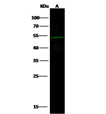
- Western Blot using ZIC3 Polyclonal Antibody (Product # PA5-81131) at 1:500 dilution. Lane A: Jurkat Whole Cell Lysate. Lysates/proteins at 30 μg per lane. Secondary Goat Anti-Rabbit IgG H&L (DyLight™ 800) at 1:10,000 dilution. Developed using the Odyssey technique. Performed under reducing conditions. Predicted band size: 54 kDa. Observed band size: 54 kDa.
Supportive validation
- Submitted by
- Invitrogen Antibodies (provider)
- Main image
- Experimental details
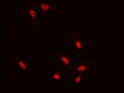
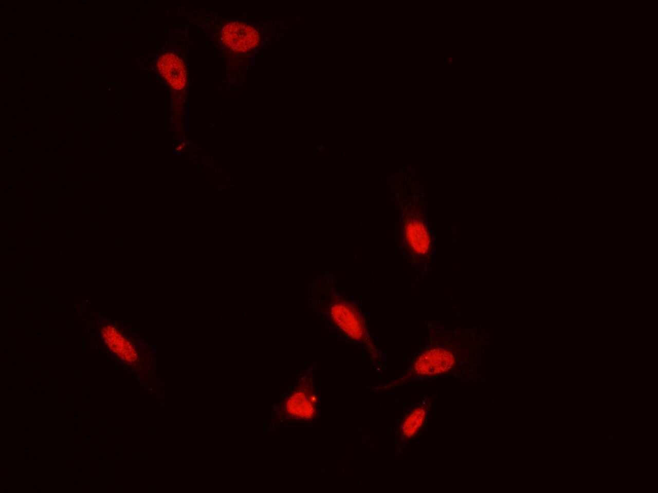
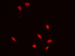
- Immunofluorescence staining of ZIC3 in HeLa cells. Cells were fixed with 4% PFA, permeabilzed with 0.3% Triton X-100 in PBS, blocked with 10% serum, and incubated with ZIC3 Polyclonal Antibody (Product # PA5-81131, 1:5,000) at 4°C overnight. Then cells were stained with the Alexa Fluor®594-conjugated Goat Anti-rabbit IgG secondary antibody (red). Positive staining was localized to cytoplasm and nucleus.
- Submitted by
- Invitrogen Antibodies (provider)
- Main image
- Experimental details
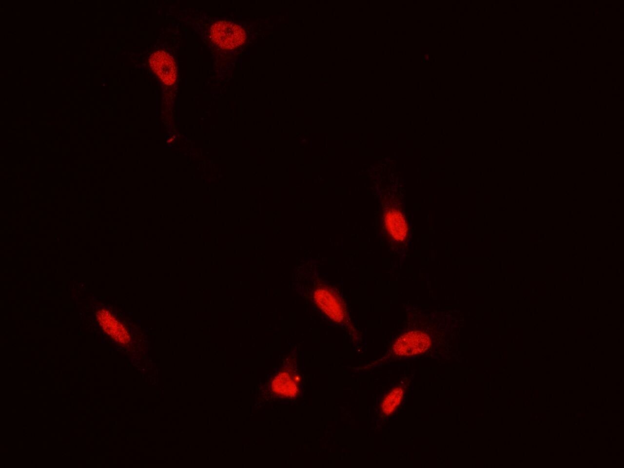
- Immunofluorescence staining of ZIC3 in HeLa cells. Cells were fixed with 4% PFA, permeabilzed with 0.3% Triton X-100 in PBS, blocked with 10% serum, and incubated with ZIC3 Polyclonal Antibody (Product # PA5-81131, 1:5,000) at 4°C overnight. Then cells were stained with the Alexa Fluor®594-conjugated Goat Anti-rabbit IgG secondary antibody (red). Positive staining was localized to cytoplasm and nucleus.
Supportive validation
- Submitted by
- Invitrogen Antibodies (provider)
- Main image
- Experimental details
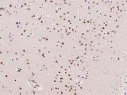
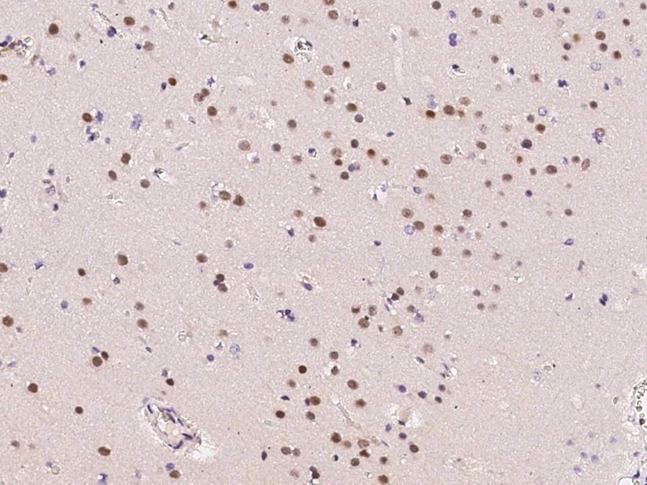
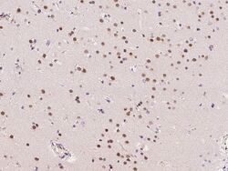
- Immunohistochemical staining of human ZIC3 in human brain with ZIC3 Polyclonal Antibody (Product # PA5-81131, 1:10,000, formalin-fixed paraffin embedded sections).
- Submitted by
- Invitrogen Antibodies (provider)
- Main image
- Experimental details
- Immunohistochemical staining of human ZIC3 in human brain with ZIC3 Polyclonal Antibody (Product # PA5-81131, 1:10,000, formalin-fixed paraffin embedded sections).

- Submitted by
- Invitrogen Antibodies (provider)
- Main image
- Experimental details
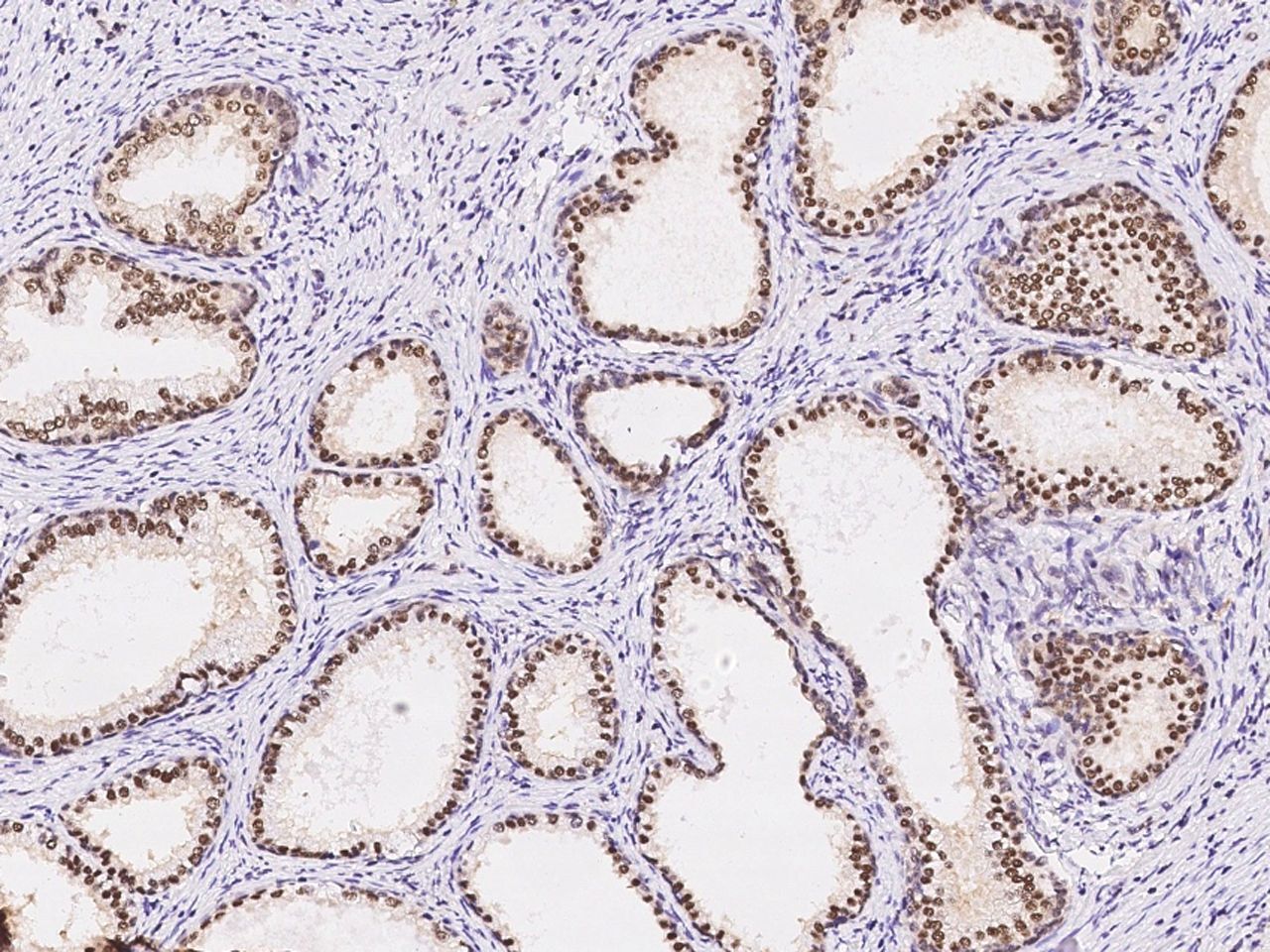
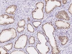
- Immunohistochemical staining of human ZIC3 in human prostate with ZIC3 Polyclonal Antibody (Product # PA5-81131, 1:10,000, formalin-fixed paraffin embedded sections).