Explore
 Validate
 Learn
Learn Immunoprecipitation
 Immunohistochemistry
Antibody data
- Antibody Data
- Antigen structure
- References [2]
- Comments [0]
- Validations
- Immunohistochemistry [1]
- Flow cytometry [1]
Submit
Validation data
Reference
Comment
Report error
- Product number
- MAB1228-100 - Provider product page

- Provider
- R&D Systems
- Product name
- Human Glycophorin A Antibody
- Antibody type
- Monoclonal
- Description
- Protein A or G purified from hybridoma culture supernatant. Detects human Glycophorin A, the major sialoglycoprotein expressed on red blood cells and erythroid precursor cells [Greaves, M.F. et al. (1983) Blood 61(4):645].
- Reactivity
- Human
- Host
- Mouse
- Conjugate
- Unconjugated
- Isotype
- IgG
- Antibody clone number
- R10
- Vial size
- 100 ug
- Storage
- Use a manual defrost freezer and avoid repeated freeze-thaw cycles. 12 months from date of receipt, -20 to -70 °C as supplied. 1 month, 2 to 8 °C under sterile conditions after reconstitution. 6 months, -20 to -70 °C under sterile conditions after reconstitution.
Submitted references Application of polychromatic flow cytometry to identify novel subsets of circulating cells with angiogenic potential.
Endothelial abnormalities in adolescents with type 1 diabetes: a biomarker for vascular sequelae?
Estes ML, Mund JA, Mead LE, Prater DN, Cai S, Wang H, Pollok KE, Murphy MP, An CS, Srour EF, Ingram DA Jr, Case J
Cytometry. Part A : the journal of the International Society for Analytical Cytology 2010 Sep;77(9):831-9
Cytometry. Part A : the journal of the International Society for Analytical Cytology 2010 Sep;77(9):831-9
Endothelial abnormalities in adolescents with type 1 diabetes: a biomarker for vascular sequelae?
DiMeglio LA, Tosh A, Saha C, Estes M, Mund J, Mead LE, Lien I, Ingram DA, Haneline LS
The Journal of pediatrics 2010 Oct;157(4):540-6
The Journal of pediatrics 2010 Oct;157(4):540-6
No comments: Submit comment
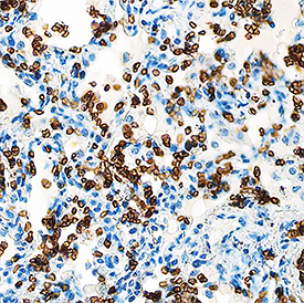
Supportive validation
- Submitted by
- R&D Systems (provider)
- Main image

- Experimental details
- Glycophorin A in Human Lung. Glycophorin A was detected in immersion fixed paraffin-embedded sections of human lung using Mouse Anti-Human Glycophorin A Monoclonal Antibody (Catalog # MAB1228) at 1.7 µg/mL overnight at 4 °C. Tissue was stained using the Anti-Mouse HRP-DAB Cell & Tissue Staining Kit (brown; Catalog # CTS002) and counterstained with hematoxylin (blue). Specific staining was localized to cell membranes of red blood cells. View our protocol for Chromogenic IHC Staining of Paraffin-embedded Tissue Sections.
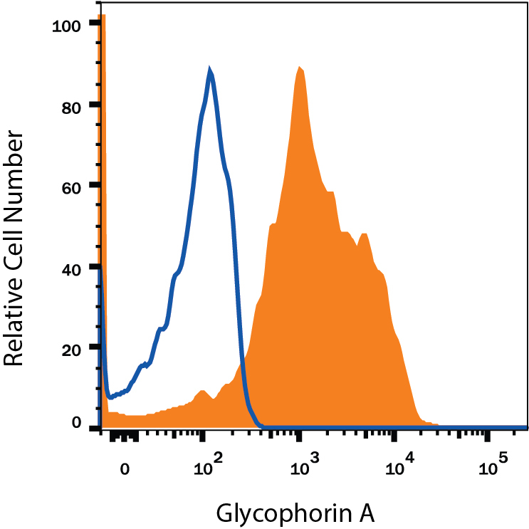
Supportive validation
- Submitted by
- R&D Systems (provider)
- Main image

- Experimental details
- Detection of Glycophorin A in TF-1 Human Cell Line by Flow Cytometry. TF-1 human erythroleukemic cell line was stained with Mouse Anti-Human Glycophorin A Monoclonal Antibody (Catalog # MAB1228, filled histogram) or isotype control antibody (Catalog # MAB002, open histogram), followed by Phycoerythrin-conjugated Anti-Mouse IgG Secondary Antibody (Catalog # F0102B).