 Explore
 Validate
 Learn
LearnNBP2-22481
antibody from Novus Biologicals
Targeting: LIN28A
CSDD1, FLJ12457, LIN-28, LIN28, ZCCHC1
 Western blot
 Immunocytochemistry Immunoprecipitation Immunohistochemistry Flow cytometry Chromatin Immunoprecipitation
Antibody data
- Antibody Data
- Antigen structure
- References [0]
- Comments [0]
- Validations
- Western blot [1]
- Immunoprecipitation [1]
- Immunohistochemistry [2]
- Flow cytometry [2]
- Chromatin Immunoprecipitation [1]
Submit
Validation data
Reference
Comment
Report error
- Product number
- NBP2-22481 - Provider product page

- Provider
- Novus Biologicals
- Product name
- Mouse Monoclonal LIN-28A Antibody
- Antibody type
- Monoclonal
- Description
- Protein A purified.
- Reactivity
- Human, Mouse
- Host
- Mouse
- Isotype
- IgG
- Vial size
- 100 ug
- Concentration
- 1 mg/ml
- Storage
- Store at -20C. Avoid freeze-thaw cycles.
No comments: Submit comment
Supportive validation
- Submitted by
- Novus Biologicals (provider)
- Main image

- Experimental details
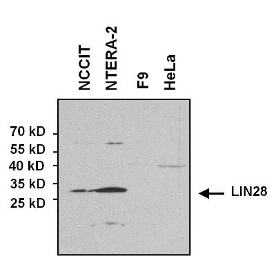
- Western Blot: LIN-28A Antibody (14E6-4E6) [NBP2-22481] - Analysis of 75 ug of various whole cell lysates and 10 ul of PageRuler Prestained Protein Ladder onto a 4-20% Tris-HCl polyacrylamide gel.
Supportive validation
- Submitted by
- Novus Biologicals (provider)
- Main image
- Experimental details
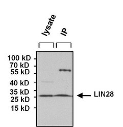
- Immunoprecipitation: LIN-28A Antibody (14E6-4E6) [NBP2-22481] - Analysis of LIN28 was performed. Antigen-antibody complexes were formed by incubating 700ug of lysate with 5 ug of an LIN28 monoclonal antibody overnight on a rocking platform at 4C. The immune complexes were captured on 50 ul Protein A/G Agarose was loaded as a positive control for detection. Samples were resolved on a 4-20% Tris-HCl polyacrylamide gel, transferred to a PVDF membrane, and blocked with 5% BSA/TBS-0.1%Tween for at least 1 hour. The membrane was probed with a LIN28 monoclonal antibody at a dilution of 1:1000 overnight rotating at 4C, washed in TBST, and probed with Clean-blot IP Detection Reagent at a dilution of 1:1000 for at least 1 hour.

Supportive validation
- Submitted by
- Novus Biologicals (provider)
- Main image

- Experimental details
- Immunohistochemistry-Paraffin: LIN-28A Antibody (14E6-4E6) [NBP2-22481] - Analysis showing staining in the nucleus and cytoplasm of mouse testis tissue (right) compared with a negative control without primary antibody (left).

- Submitted by
- Novus Biologicals (provider)
- Main image

- Experimental details
- Immunohistochemistry-Paraffin: LIN-28A Antibody (14E6-4E6) [NBP2-22481] - Analysis showing staining in the cytoplasm of human seminoma (right) compared with a negative control without primary antibody (left).

Supportive validation
- Submitted by
- Novus Biologicals (provider)
- Main image
- Experimental details
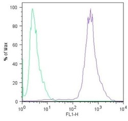
- Flow Cytometry: LIN-28A Antibody (14E6-4E6) [NBP2-22481] - Analysis of Lin28 (blue histogram) on HEL 11.4 induced IPS cells. To generate single cells suspensions, colonies were treated with TrypLE cell dissociation enzyme for 5 minutes at 37C. Cells were incubated with a Lin28 monoclonal antibody or mouse IgG (green histogram) at a dilution of 1:100 for 1 hour on ice, washed with PBS + 5% fetal calf serum (FACS buffer), and incubated with a fluorescein-conjugated secondary antibody at a dilution of 1:200 for 30 minutes on ice. Cells were washed with cold FACS buffer, resuspended in 500ul of FACS buffer containing 10ul of 4% paraformaldehyde.

- Submitted by
- Novus Biologicals (provider)
- Main image

- Experimental details
- Flow Cytometry: LIN-28A Antibody (14E6-4E6) [NBP2-22481] - Analysis of Lin28 (blue histogram) on H9 embryonic stem cells. To generate single cells suspensions, colonies were treated with TrypLE cell dissociation enzyme for 5 minutes at 37C. Cells were incubated with a Lin28 monoclonal antibody or mouse IgG (green histogram) at a dilution of 1:100 for 1 hour on ice, washed with PBS + 5% fetal calf serum (FACS buffer), and incubated with a fluorescein-conjugated secondary antibody at a dilution of 1:200 for 30 minutes on ice. Cells were washed with cold FACS buffer, resuspended in 500ul of FACS buffer containing 10ul of 4% paraformaldehyde.

Supportive validation
- Submitted by
- Novus Biologicals (provider)
- Main image

- Experimental details
- Chromatin Immunoprecipitation: LIN-28A Antibody (14E6-4E6) [NBP2-22481] - Analysis performed using cross-linked chromatin from rat hepatoma cells treated with insulin. IP performed using a multiplex microplate Matrix ChIP assay of LIN28 monoclonal antibody. Chromatin aliquots from cells were used per ChIP pull-down. Quantitative PCR data done in quadruplicate using 1ul of DNA in 2ul SYBR real-time PCR reactions containing primers to amplify -15kb upstream of Egr1 or exon-1 or exon-2-3 of Egr1. Quantitation of immunoprecipitated chromatin is presented as signal relative to the total amount of input chromatin. Results represent the mean +/- SEM. A schematic representation of the rat Egr-1 locus is shown; oxes represent exons (black boxes = translated, white boxes = untranslated), the zigzag line represents an intron, and the straight line represents upstream sequence. Regions amplified by Egr-1 primers are represented by black bars. Data courtesy of the Innovators Program.
