 Explore
 Validate
 Learn
Learn Western blot
Antibody data
- Antibody Data
- Antigen structure
- References [10]
- Comments [0]
- Validations
- Western blot [1]
- Immunocytochemistry [2]
Submit
Validation data
Reference
Comment
Report error
- Product number
- AF3757 - Provider product page

- Provider
- R&D Systems
- Product name
- Human LIN-28A Antibody
- Antibody type
- Polyclonal
- Description
- Immunogen affinity purified. Detects human LIN-28A in direct ELISAs and Western blots.
- Reactivity
- Human
- Host
- Goat
- Conjugate
- Unconjugated
- Antigen sequence
Q9H9Z2- Isotype
- IgG
- Vial size
- 100 ug
- Concentration
- LYOPH
- Storage
- Use a manual defrost freezer and avoid repeated freeze-thaw cycles. 12 months from date of receipt, -20 to -70 °C as supplied. 1 month, 2 to 8 °C under sterile conditions after reconstitution. 6 months, -20 to -70 °C under sterile conditions after reconstitution.
Submitted references Exonuclease Domain-Containing 1 Enhances MIWI2 piRNA Biogenesis via Its Interaction with TDRD12.
Autologous and Heterologous Cell Therapy for Hemophilia B toward Functional Restoration of Factor IX.
Biallelic loss of human CTNNA2, encoding αN-catenin, leads to ARP2/3 complex overactivity and disordered cortical neuronal migration.
Derivation of induced pluripotent stem cells in Japanese macaque (Macaca fuscata).
Protein-driven RNA nanostructured devices that function in vitro and control mammalian cell fate.
Glycolysis-Optimized Conditions Enhance Maintenance of Regenerative Integrity in Mouse Spermatogonial Stem Cells during Long-Term Culture.
Embryonic lethality and defective male germ cell development in mice lacking UTF1.
Reconstruction of mouse testicular cellular microenvironments in long-term seminiferous tubule culture.
Activation of pluripotency genes in human fibroblast cells by a novel mRNA based approach.
Induction of pluripotent stem cells from adult human fibroblasts by defined factors.
Pandey RR, Homolka D, Olotu O, Sachidanandam R, Kotaja N, Pillai RS
Cell reports 2018 Sep 25;24(13):3423-3432.e4
Cell reports 2018 Sep 25;24(13):3423-3432.e4
Autologous and Heterologous Cell Therapy for Hemophilia B toward Functional Restoration of Factor IX.
Ramaswamy S, Tonnu N, Menon T, Lewis BM, Green KT, Wampler D, Monahan PE, Verma IM
Cell reports 2018 May 1;23(5):1565-1580
Cell reports 2018 May 1;23(5):1565-1580
Biallelic loss of human CTNNA2, encoding αN-catenin, leads to ARP2/3 complex overactivity and disordered cortical neuronal migration.
Schaffer AE, Breuss MW, Caglayan AO, Al-Sanaa N, Al-Abdulwahed HY, Kaymakçalan H, Yılmaz C, Zaki MS, Rosti RO, Copeland B, Baek ST, Musaev D, Scott EC, Ben-Omran T, Kariminejad A, Kayserili H, Mojahedi F, Kara M, Cai N, Silhavy JL, Elsharif S, Fenercioglu E, Barshop BA, Kara B, Wang R, Stanley V, James KN, Nachnani R, Kalur A, Megahed H, Incecik F, Danda S, Alanay Y, Faqeih E, Melikishvili G, Mansour L, Miller I, Sukhudyan B, Chelly J, Dobyns WB, Bilguvar K, Jamra RA, Gunel M, Gleeson JG
Nature genetics 2018 Aug;50(8):1093-1101
Nature genetics 2018 Aug;50(8):1093-1101
Derivation of induced pluripotent stem cells in Japanese macaque (Macaca fuscata).
Nakai R, Ohnuki M, Kuroki K, Ito H, Hirai H, Kitajima R, Fujimoto T, Nakagawa M, Enard W, Imamura M
Scientific reports 2018 Aug 15;8(1):12187
Scientific reports 2018 Aug 15;8(1):12187
Protein-driven RNA nanostructured devices that function in vitro and control mammalian cell fate.
Shibata T, Fujita Y, Ohno H, Suzuki Y, Hayashi K, Komatsu KR, Kawasaki S, Hidaka K, Yonehara S, Sugiyama H, Endo M, Saito H
Nature communications 2017 Sep 14;8(1):540
Nature communications 2017 Sep 14;8(1):540
Glycolysis-Optimized Conditions Enhance Maintenance of Regenerative Integrity in Mouse Spermatogonial Stem Cells during Long-Term Culture.
Helsel AR, Oatley MJ, Oatley JM
Stem cell reports 2017 May 9;8(5):1430-1441
Stem cell reports 2017 May 9;8(5):1430-1441
Embryonic lethality and defective male germ cell development in mice lacking UTF1.
Kasowitz SD, Luo M, Ma J, Leu NA, Wang PJ
Scientific reports 2017 Dec 8;7(1):17259
Scientific reports 2017 Dec 8;7(1):17259
Reconstruction of mouse testicular cellular microenvironments in long-term seminiferous tubule culture.
Mäkelä JA, Toppari J, Rivero-Müller A, Ventelä S
PloS one 2014;9(3):e90088
PloS one 2014;9(3):e90088
Activation of pluripotency genes in human fibroblast cells by a novel mRNA based approach.
Plews JR, Li J, Jones M, Moore HD, Mason C, Andrews PW, Na J
PloS one 2010 Dec 30;5(12):e14397
PloS one 2010 Dec 30;5(12):e14397
Induction of pluripotent stem cells from adult human fibroblasts by defined factors.
Takahashi K, Tanabe K, Ohnuki M, Narita M, Ichisaka T, Tomoda K, Yamanaka S
Cell 2007 Nov 30;131(5):861-72
Cell 2007 Nov 30;131(5):861-72
No comments: Submit comment
Supportive validation
- Submitted by
- R&D Systems (provider)
- Main image
- Experimental details
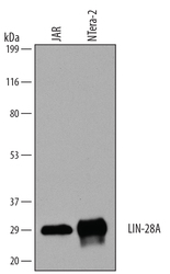
- Detection of Human LIN-28A by Western Blot. Western blot shows lysates of JAR human choriocarcinoma cell line and NTera-2 human testicular embryonic carcinoma cell line. PVDF membrane was probed with 0.1 µg/mL of Goat Anti-Human LIN-28A Antigen Affinity-purified Polyclonal Antibody (Catalog # AF3757) followed by HRP-conjugated Anti-Goat IgG Secondary Antibody (Catalog # HAF109). A specific band was detected for LIN-28A at approximately 30 kDa (as indicated). This experiment was conducted under reducing conditions and using Immunoblot Buffer Group 1.

Supportive validation
- Submitted by
- R&D Systems (provider)
- Main image
- Experimental details
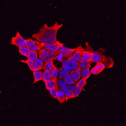
- LIN-28A in D3 Mouse Stem Cells. LIN-28A was detected in immersion fixed D3 mouse embryonic stem cell line using Goat Anti-Human LIN-28A Antigen Affinity-purified Polyclonal Antibody (Catalog # AF3757) at 10 µg/mL for 3 hours at room temperature. Cells were stained using the Northern-Lights™ 557-conjugated Anti-Goat IgG Secondary Antibody (red; Catalog # NL001) and counterstained with DAPI (blue). Specific staining was localized to cytoplasm. View our protocol for Fluorescent ICC Staining of Cells on Coverslips.

- Submitted by
- R&D Systems (provider)
- Main image
- Experimental details
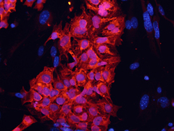
- LIN-28A in BG01V Human Stem Cells. LIN-28A was detected in immersion fixed BG01V human embryonic stem cells using 10 µg/mL Goat Anti-Human LIN-28A Antigen Affinity-purified Polyclonal Antibody (Catalog # AF3757) for 3 hours at room temperature. Cells were stained with the NorthernLights™ 557-conjugated Anti-Goat IgG Secondary Antibody (red; Catalog # NL001) and counter-stained with DAPI (blue). View our protocol for Fluorescent ICC Staining of Cells on Coverslips.
