 Explore
 Validate
 Learn
Learn Western blot
 Immunohistochemistry
Antibody data
- Antibody Data
- Antigen structure
- References [0]
- Comments [0]
- Validations
- Western blot [2]
- Immunocytochemistry [1]
- Chromatin Immunoprecipitation [1]
Submit
Validation data
Reference
Comment
Report error
- Product number
- ABIN2616404 - Provider product page

- Provider
- antibodies-online
- Product name
- anti-Anoctamin 1, Calcium Activated Chloride Channel (ANO1) antibody
- Antibody type
- Monoclonal
- Description
- Tissue culture supernatant
- Reactivity
- Human
- Host
- Rabbit
- Isotype
- IgG
- Antibody clone number
- RBT-DOG1
- Vial size
- 500 μL
- Storage
- Store at 2°C to 8°C.
No comments: Submit comment
Supportive validation
- Submitted by
- antibodies-online (provider)
- Main image
- Experimental details
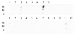
- Histone H3 dimethyl Lys9 antibody tested by dot blot analysis. Dot blot analysis was used to confirm the specificity of Histone H3 dimethyl Lys9 antibody for dimethyl Lys9 histone H3. Methylated peptides corresponding to the immunogen and related sequences derived from histone H3 were spotted onto PVDF and probed with the antibody at 1:10,000. The amount of peptide (picomoles) spotted is indicated next to each row. Top row:Lane 1: Unmod H3 aa 1-10. Lane 2: monomethyl Lys4. Lane 3: dimethyl Lys4. Lane 4: trimethyl Lys4. Lane 5: Unmod H3 aa 6-22. Lane 6: monomethyl Lys9. Lane 7: dimethyl Lys9. Lane 8: trimethyl Lys9. Bottom row: Lane 1: dimethyl Lys14. Lane 2: monomethyl Lys18. Lane 3: dimethyl Lys18. Lane 4: trimethyl Lys18. Lane 5: unmodified corresponding to amino acids 18-27 of human histone H3. Lane 6: monomethyl Lys23. Lane 7: dimethyl Lys23. Lane 8: trimethyl Lys23. Lane 9: unmodified corresponding to amino acids 22-32 of human histone H3. Lane 10: monomethyl Lys27. Lane 11: dimethyl Lys27. Lane 12: trimethyl Lys27.

- Submitted by
- antibodies-online (provider)
- Main image

- Experimental details
- Histone H3 dimethyl Lys9 antibody tested by Western blot. HeLa acid extract (10 μg per lane) probed with Histone H3 dimethyl Lys9 antibody at a 1:10,000 dilution.

Supportive validation
- Submitted by
- antibodies-online (provider)
- Main image

- Experimental details
- Histone H3 dimethyl Lys9 antibody tested by immunofluorescence. Top left: HeLa cells stained with Histone H3 dimethyl Lys9 antibody (1:1,000). Top right: Same cells stained with alpha Tubulin mAb (Clone 5-B-1-2). Bottom left: Same cells stained with DAPI. Bottom right: Merge of all 3 images.

Supportive validation
- Submitted by
- antibodies-online (provider)
- Main image

- Experimental details
- Histone H3 dimethyl Lys9 antibody tested by ChIP. Chromatin IP performed using the ChIP-IT® Express Kit (Catalog No. 53008) and HeLa Chromatin (1.5 x 10⁶ cell equivalents per ChIP) using 10 μl of Histone H3 dimethyl Lys9 antibody or the equivalent amount of rabbit IgG as a negative control. Real time, quantitative PCR (RT-qPCR) was performed on DNA purified from each of the ChIP reactions using a primer pair specific for either the PABPC1 gene or the MyoD gene. Data are presented as Fold Enrichment of the ChIP antibody signal versus the negative control IgG using the ddCT method.
