 Explore
 Validate
 Learn
Learn Western blot
Antibody data
- Antibody Data
- Antigen structure
- References [0]
- Comments [0]
- Validations
- Western blot [4]
- Immunocytochemistry [1]
Submit
Validation data
Reference
Comment
Report error
- Product number
- MAB85582-100 - Provider product page

- Provider
- R&D Systems
- Product name
- Human/Mouse/Rat LC3B Antibody
- Antibody type
- Monoclonal
- Description
- Protein A or G purified from cell culture supernatant. Detects human, mouse, and rat LC3B in Western blots.
- Reactivity
- Human, Mouse, Rat
- Host
- Rabbit
- Conjugate
- Unconjugated
- Antigen sequence
Q9GZQ8- Isotype
- IgG
- Antibody clone number
- 1251B
- Vial size
- 100 ug
- Storage
- Use a manual defrost freezer and avoid repeated freeze-thaw cycles. 12 months from date of receipt, -20 to -70 °C as supplied. 1 month, 2 to 8 °C under sterile conditions after reconstitution. 6 months, -20 to -70 °C under sterile conditions after reconstitution.
No comments: Submit comment
Supportive validation
- Submitted by
- R&D Systems (provider)
- Main image

- Experimental details
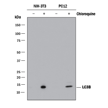
- Detection of Mouse and Rat LC3B by Western Blot. Western blot shows lysates of NIH-3T3 mouse embryonic fibroblast cell line and PC-12 rat adrenal pheochromocytoma cell line untreated (-) or treated (+) with 50uM Chloroquine for 18 hours. PVDF membrane was probed with 0.1 µg/mL of Rabbit Anti-Human/Mouse/Rat LC3B Monoclonal Antibody (Catalog # MAB85582) followed by HRP-conjugated Anti-Rabbit IgG Secondary Antibody (Catalog # HAF008). A specific band was detected for LC3B at approximately 15 kDa (as indicated). This experiment was conducted under reducing conditions and using Immunoblot Buffer Group 1.
- Submitted by
- R&D Systems (provider)
- Main image

- Experimental details
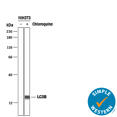
- Detection of Mouse LC3B by Simple WesternTM. Simple Western lane view shows lysates of NIH-3T3 mouse embryonic fibroblast cell line untreated (-) or treated (+) with 50uM Chloroquine for 18 hours, loaded at 0.2 mg/mL. A specific band was detected for LC3B at approximately 17 kDa (as indicated) using 5 µg/mL of Rabbit Anti-Human/Mouse/Rat LC3B Monoclonal Antibody (Catalog # MAB85582) . This experiment was conducted under reducing conditions and using the 12-230 kDa separation system.
- Submitted by
- R&D Systems (provider)
- Main image
- Experimental details
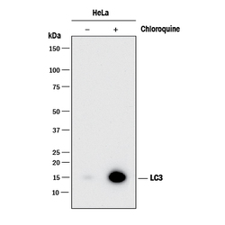
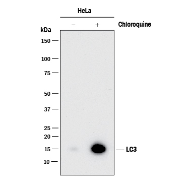
- Detection of Human LC3B by Western Blot. Western blot shows lysates of HeLa human cervical epithelial carcinoma cell line untreated (-) or treated (+) with 50uM Chloroquine for 18 Hours. PVDF membrane was probed with 0.1 µg/mL of Rabbit Anti-Human/Mouse/Rat LC3B Monoclonal Antibody (Catalog # MAB85582) followed by HRP-conjugated Anti-Rabbit IgG Secondary Antibody (Catalog # HAF008). A specific band was detected for LC3B at approximately 15 kDa (as indicated). This experiment was conducted under reducing conditions and using Immunoblot Buffer Group 1.
- Submitted by
- R&D Systems (provider)
- Main image
- Experimental details
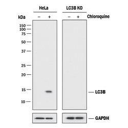
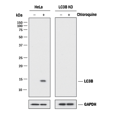
- Western Blow Show Human LC3B Specificty Using Knockout Cell Line. Western blot shows lysates of HeLa human cervical epithelial carcinoma parental cell line and LC3B knockout HeLa cell line (KO) untreated (-) or treated (+) with 50uM Chloroquine for 18 hours. PVDF membrane was probed with 0.1 µg/mL of Rabbit Anti-Human/Mouse/Rat LC3B Monoclonal Antibody (Catalog # MAB85582) followed by HRP-conjugated Anti-Rabbit IgG Secondary Antibody (Catalog # HAF008). A specific band was detected for LC3B at approximately 15 kDa (as indicated) in the parental HeLa cell line, but is not detectable in the knockout HeLa cell line. GAPDH (Catalog # AF5718) is shown as a loading control. This experiment was conducted under reducing conditions and using Immunoblot Buffer Group 1.
Supportive validation
- Submitted by
- R&D Systems (provider)
- Main image
- Experimental details
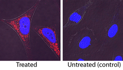
- LC3B in HeLa Human Cell Line. LC3B was detected in immersion fixed HeLa human cervical epithelial carcinoma cell line treated with Chloroquine using Rabbit Anti-Human/Mouse/Rat LC3B Monoclonal Antibody (Catalog # MAB85582) at 1 µg/mL for 3 hours at room temperature. Cells were stained using the NorthernLights™ 557-conjugated Anti-Rabbit IgG Secondary Antibody (red; Catalog # NL004) and counterstained with DAPI (blue). Specific staining was localized to cytoplasm. View our protocol for Fluorescent ICC Staining of Cells on Coverslips.
