Explore
 Validate
 Learn
Learn Western blot
Antibody data
- Antibody Data
- Antigen structure
- References [0]
- Comments [0]
- Validations
- Western blot [3]
- Immunocytochemistry [2]
- Immunohistochemistry [2]
Submit
Validation data
Reference
Comment
Report error
- Product number
- AP32166PU-N - Provider product page

- Provider
- Acris Antibodies GmbH
- Product name
- anti LC3B (N-term)
- Antibody type
- Polyclonal
- Antigen
- KLH conjugated synthetic peptide between 1~30 amino acids from the N-terminal region of Human LC3 (APG8b)
- Reactivity
- Human, Rat
- Host
- Rabbit
- Vial size
- 0.4 ml
- Concentration
- lot specific
No comments: Submit comment
Supportive validation
- Submitted by
- Acris Antibodies GmbH (provider)
- Main image

- Experimental details
- Western blot analysis of LC3B Antibody Cat.-No AP32166PU-N in 293 cell line lysates transiently transfected with the LC3 (APG8b) gene (2 µg/lane). Human LC3 (APG8b) (arrow) was detected using the purified Pab (1/60 dilution).

- Submitted by
- Acris Antibodies GmbH (provider)
- Main image
- Experimental details
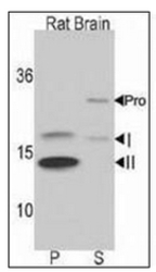
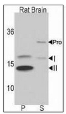
- Western blot analysis of LC3B Antibody Cat.-No AP32166PU-NÂ in Rat brain lysate. Both non-lipidated (arrow, I) and lipidated LC3 (APG8b) (arrow, II) were detected in membrane fraction (P) but pro-LC3 (APG8b) and non-lipidated LC3 (APG8b) were detected in soluble fraction (S).
- Submitted by
- Acris Antibodies GmbH (provider)
- Main image
- Experimental details
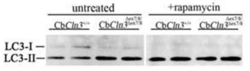
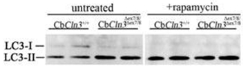
- Immunoblots for LC3 protein (LC3B Antibody Cat.-No AP32166PU-N) are shown for extracts from untreated or rapamycin-treated (250 nM, 4 h) cultures of wild-type (CbCln3+/+) or homozygous cerebellar cells (CbCln3Ãex7/8/Ãex7/8). Rapamycin increases the LC3-II/LC3-I ratio in wild-type and homogyzous cerebellar cell extracts (2.6 and 2.2-fold, respectively), consistent with activation of autophagy.
Supportive validation
- Submitted by
- Acris Antibodies GmbH (provider)
- Main image
- Experimental details
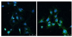
- Immunofluorescence analysis of LC3B Antibody Cat.-No AP32166PU-NÂ in Rat brain before (left) and after (right) treatment with rapamycin (100 nm for 4 hr).

- Submitted by
- Acris Antibodies GmbH (provider)
- Main image
- Experimental details
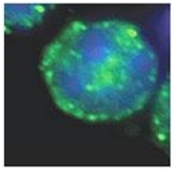
- FACS sorted LSK cells (Lineage-Sca-1+c-kit+, enriched for hematopoietic stem cells) incubated with LC3B Antibody Cat.-No AP32166PU-N at 1/50 dilution at 4 degree overnight., and detected with Alexa Fluor 488 conjugated anti-Rabbit antibody (1/200). Data courtesy of Wenbin Xiao, La Jolla Institute for Allergy and Immunology.

Supportive validation
- Submitted by
- Acris Antibodies GmbH (provider)
- Main image

- Experimental details
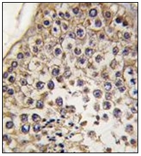
- Formalin-Fixed, Paraffin-Embedded human testis tissue reacted with Autophagy LC3B Antibody Cat.-No AP32166PU-N, which was peroxidase-conjugated to the secondary antibody, followed by DAB staining.
- Submitted by
- Acris Antibodies GmbH (provider)
- Main image
- Experimental details
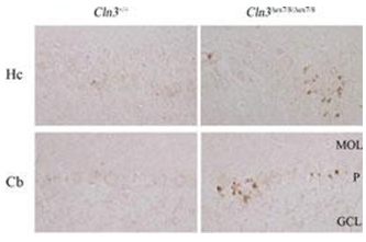
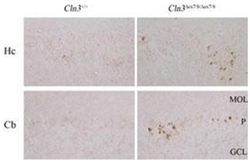
- Wild-type (Cln3+/+) or homozygous Cln3ex7/8 (Cln3ex7/8/ex7/8) paraffin-embedded brain sections immunostained for the LC3 protein (Autophagy LC3B Antibody Cat.-No AP32166PU-N). Shown are the CA2/CA3 region of hippocampus (Hc) and cerebellum (Cb) from 10-month-old mice. Few immunopositive puncta are present in wild-type sections, whereas homozygous Cln3ex7/8 sections contain clusters of LC3-positive puncta around pyramidal neurons and Purkinje cells (P). MOL, molecular layer; GCL, granule cell layer. Data courtesy of Dr. Susan Cotman, Massachusets General Hospital.