 Explore
 Validate
 Learn
Learn Western blot
 Immunocytochemistry
Antibody data
- Antibody Data
- Antigen structure
- References [1]
- Comments [0]
- Validations
- Western blot [2]
- Immunohistochemistry [7]
- Flow cytometry [2]
Submit
Validation data
Reference
Comment
Report error
- Product number
- NBP2-01800 - Provider product page

- Provider
- Novus Biologicals
- Proper citation
- Novus Cat#NBP2-01800, RRID:AB_2725750
- Product name
- Mouse Monoclonal Cytochrome P450 2B6 Antibody
- Antibody type
- Monoclonal
- Description
- Affinity purified.
- Reactivity
- Human, Mouse, Rat, Canine, Simian
- Host
- Mouse
- Isotype
- IgG
- Vial size
- 0.1 ml
- Concentration
- 1.1 mg/ml
- Storage
- Store at -20C. Avoid freeze-thaw cycles.
Submitted references Xenobiotic Nuclear Receptor Signaling Determines Molecular Pathogenesis of Progressive Familial Intrahepatic Cholestasis.
Kim KH, Choi JM, Li F, Arizpe A, Wooton-Kee CR, Anakk S, Jung SY, Finegold MJ, Moore DD
Endocrinology 2018 Jun 1;159(6):2435-2446
Endocrinology 2018 Jun 1;159(6):2435-2446
No comments: Submit comment
Supportive validation
- Submitted by
- Novus Biologicals (provider)
- Main image

- Experimental details
- Western Blot: Cytochrome P450 2B6 Antibody (3D5) [NBP2-01800] - HEK293T cells were transfected with the pCMV6-ENTRY control (Left lane) or pCMV6-ENTRY Cytochrome P450 2B6 (Right lane) cDNA for 48 hrs and lysed. Equivalent amounts of cell lysates (5 ug per lane) were separated by SDS-PAGE and immunoblotted with anti-Cytochrome P450 2B6.

- Submitted by
- Novus Biologicals (provider)
- Main image
- Experimental details
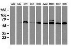
- Western Blot: Cytochrome P450 2B6 Antibody (3D5) [NBP2-01800] - Analysis of extracts (35ug) from 9 different cell lines by using anti-Cytochrome P450 2B6 monoclonal antibody (HepG2: human; HeLa: human; SVT2: mouse; A549: human; COS7: monkey; Jurkat: human; MDCK: canine; PC12: rat; MCF7: human).

Supportive validation
- Submitted by
- Novus Biologicals (provider)
- Main image

- Experimental details
- Immunohistochemistry-Paraffin: Cytochrome P450 2B6 Antibody (3D5) [NBP2-01800] - Staining of paraffin-embedded Human liver tissue using anti-Cytochrome P450 2B6 mouse monoclonal antibody.

- Submitted by
- Novus Biologicals (provider)
- Main image
- Experimental details
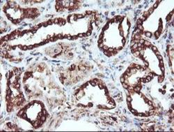
- Immunohistochemistry-Paraffin: Cytochrome P450 2B6 Antibody (3D5) [NBP2-01800] - Staining of paraffin-embedded Human Kidney tissue using anti-Cytochrome P450 2B6 mouse monoclonal antibody.

- Submitted by
- Novus Biologicals (provider)
- Main image

- Experimental details
- Immunohistochemistry-Paraffin: Cytochrome P450 2B6 Antibody (3D5) [NBP2-01800] - Staining of paraffin-embedded Human colon tissue using anti-Cytochrome P450 2B6 mouse monoclonal antibody.

- Submitted by
- Novus Biologicals (provider)
- Main image
- Experimental details
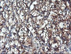
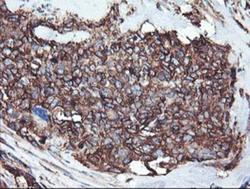
- Immunohistochemistry-Paraffin: Cytochrome P450 2B6 Antibody (3D5) [NBP2-01800] - Staining of paraffin-embedded Carcinoma of Human kidney tissue using anti-Cytochrome P450 2B6 mouse monoclonal antibody.
- Submitted by
- Novus Biologicals (provider)
- Main image

- Experimental details
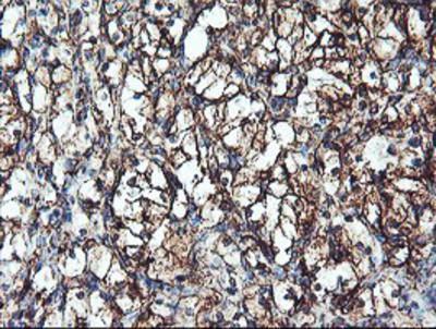
- Immunohistochemistry-Paraffin: Cytochrome P450 2B6 Antibody (3D5) [NBP2-01800] - Staining of paraffin-embedded Carcinoma of Human bladder tissue using anti-Cytochrome P450 2B6 mouse monoclonal antibody.

- Submitted by
- Novus Biologicals (provider)
- Main image

- Experimental details
- Immunohistochemistry-Paraffin: Cytochrome P450 2B6 Antibody (3D5) [NBP2-01800] - Staining of paraffin-embedded Adenocarcinoma of Human colon tissue using anti-Cytochrome P450 2B6 mouse monoclonal antibody.

- Submitted by
- Novus Biologicals (provider)
- Main image
- Experimental details
- Immunohistochemistry-Paraffin: Cytochrome P450 2B6 Antibody (3D5) [NBP2-01800] - Staining of paraffin-embedded Adenocarcinoma of Human breast tissue using anti-Cytochrome P450 2B6 mouse monoclonal antibody.

Supportive validation
- Submitted by
- Novus Biologicals (provider)
- Main image
- Experimental details
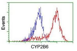
- Flow Cytometry: Cytochrome P450 2B6 Antibody (3D5) [NBP2-01800] - HEK293T cells transfected with either overexpression plasmid (Red) or empty vector control plasmid (Blue) were immunostained by anti-Cytochrome P450 2B6 antibody, and then analyzed by flow cytometry.

- Submitted by
- Novus Biologicals (provider)
- Main image
- Experimental details
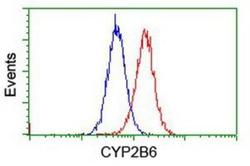
- Flow Cytometry: Cytochrome P450 2B6 Antibody (3D5) [NBP2-01800] - Analysis of Jurkat cells, using anti-Cytochrome P450 2B6 antibody, (Red), compared to a nonspecific negative control antibody (Blue).
