Explore
 Validate
 Learn
Learn Western blot
Antibody data
- Antibody Data
- Antigen structure
- References [3]
- Comments [0]
- Validations
- Western blot [3]
- Immunocytochemistry [2]
- Immunohistochemistry [1]
Submit
Validation data
Reference
Comment
Report error
- Product number
- GTX113727 - Provider product page

- Provider
- GeneTex
- Proper citation
- GeneTex Cat#GTX113727, RRID:AB_2037134
- Product name
- HADHA antibody [N2C1], Internal
- Antibody type
- Polyclonal
- Reactivity
- Human, Mouse, Rat
- Host
- Rabbit
Submitted references Hepatic Fatty Acid Oxidation Restrains Systemic Catabolism during Starvation.
Adipose fatty acid oxidation is required for thermogenesis and potentiates oxidative stress-induced inflammation.
Chemical-genetic induction of Malonyl-CoA decarboxylase in skeletal muscle.
Lee J, Choi J, Scafidi S, Wolfgang MJ
Cell reports 2016 Jun 28;16(1):201-212
Cell reports 2016 Jun 28;16(1):201-212
Adipose fatty acid oxidation is required for thermogenesis and potentiates oxidative stress-induced inflammation.
Lee J, Ellis JM, Wolfgang MJ
Cell reports 2015 Jan 13;10(2):266-79
Cell reports 2015 Jan 13;10(2):266-79
Chemical-genetic induction of Malonyl-CoA decarboxylase in skeletal muscle.
Rodriguez S, Ellis JM, Wolfgang MJ
BMC biochemistry 2014 Aug 25;15:20
BMC biochemistry 2014 Aug 25;15:20
No comments: Submit comment
Supportive validation
- Submitted by
- GeneTex (provider)
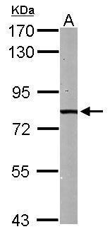
- Main image

- Experimental details
- Sample (50 ug of whole cell lysate) A: mouse liver 7.5% SDS PAGE GTX113727 diluted at 1:1000
- Submitted by
- GeneTex (provider)
- Main image

- Experimental details
- HADHA antibody [N2C1], Internal detects HADHA protein by Western blot analysis.A.30 ?g PC-12 whole cell lysate/extract 7.5 % SDS-PAGEHADHA antibody [N2C1], Internal (GTX113727) dilution: 1:500

- Submitted by
- GeneTex (provider)
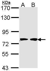
- Main image

- Experimental details
- Sample (30 ug of whole cell lysate) A: 293T B: A431 (GTX27909) 7.5% SDS PAGE GTX113727 diluted at 1:1000
Supportive validation
- Submitted by
- GeneTex (provider)
- Main image
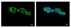
- Experimental details
- HADHA antibody [N2C1], Internal detects HADHA protein at Mitochondia by immunofluorescent analysis. Sample: HepG2 cells were fixed in -20¢J 100% MeOH for 5 min.Green: HADHA protein stained by HADHA antibody [N2C1], Internal (GTX113727) diluted at 1:500.Blue: Hoechst 33343 staining.

- Submitted by
- GeneTex (provider)
- Main image

- Experimental details
- HADHA antibody [N2C1], Internal detects HADHA protein at cytoplasm by immunofluorescent analysis.Sample: HeLa cells were fixed in 4% paraformaldehyde at RT for 15 min.Green: HADHA protein stained by HADHA antibody [N2C1], Internal (GTX113727) diluted at 1:500.Red: alpha Tubulin, a cytoskeleton marker, stained by alpha Tubulin antibody [GT114] (GTX628802) diluted at 1:1000.Blue: Hoechst 33342 staining.

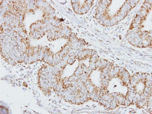
Supportive validation
- Submitted by
- GeneTex (provider)
- Main image

- Experimental details
- Immunohistochemical analysis of paraffin-embedded human colon carcinoma, using HADHA(GTX113727) antibody at 1:250 dilution.