 Explore
 Validate
 Learn
Learn Western blot
Antibody data
- Antibody Data
- Antigen structure
- References [1]
- Comments [0]
- Validations
- Western blot [5]
- Immunocytochemistry [1]
- Immunohistochemistry [1]
- Other assay [1]
Submit
Validation data
Reference
Comment
Report error
- Product number
- PA5-27348 - Provider product page

- Provider
- Invitrogen Antibodies
- Product name
- HADHA Polyclonal Antibody
- Antibody type
- Polyclonal
- Antigen
- Recombinant protein fragment
- Reactivity
- Human, Mouse, Rat
- Host
- Rabbit
- Isotype
- IgG
- Vial size
- 100 µL
- Concentration
- 1 mg/mL
- Storage
- Store at 4°C short term. For long term storage, store at -20°C, avoiding freeze/thaw cycles.
Submitted references PKA Activates AMPK Through LKB1 Signaling in Follicular Thyroid Cancer.
Kari S, Vasko VV, Priya S, Kirschner LS
Frontiers in endocrinology 2019;10:769
Frontiers in endocrinology 2019;10:769
No comments: Submit comment
Supportive validation
- Submitted by
- Invitrogen Antibodies (provider)
- Main image

- Experimental details
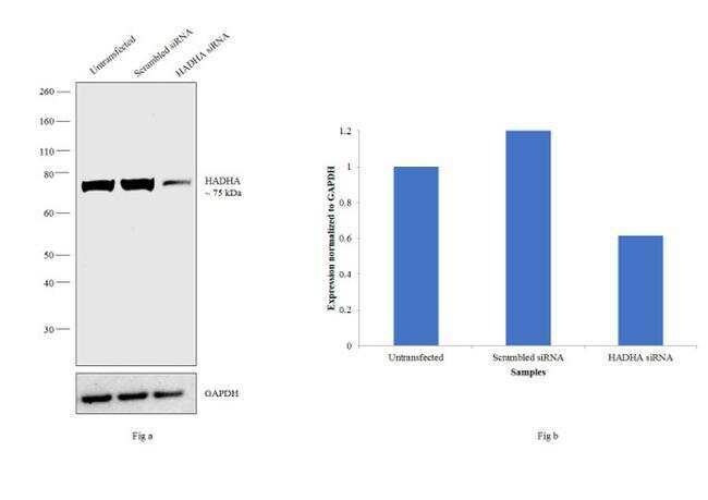
- Knockdown of HADHA was achieved by transfecting Hep G2 with HADHA specific siRNAs (Silencer® select Product # s6441). Western blot analysis (Fig. a) was performed using membrane enriched extracts from the HADHA knockdown cells (lane 3), non-specific scrambled siRNA transfected cells (lane 2) and untransfected cells (lane 1). The blot was probed with HADHA Polyclonal Antibody (Product # PA5-27348, 1:1000 dilution) and Goat anti-Rabbit IgG (H+L) Superclonal™ Secondary Antibody, HRP conjugate (Product # A27036, 0.25µg/ml, 1:4000 dilution). Densitometric analysis of this western blot is shown in histogram (Fig. b). Decrease in signal upon siRNA mediated knock down confirms that antibody is specific to HADHA.
- Submitted by
- Invitrogen Antibodies (provider)
- Main image

- Experimental details
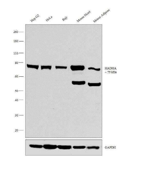
- Western blot analysis was performed on membrane enriched extracts (30 µg lysate) of Hep G2 (Lane 1), HeLa (Lane 2), Raji (Lane 3), tissue extracts of Mouse Heart (Lane 4) and Mouse Adipose (Lane 5). The blot was probed with Anti-HADHA Polyclonal Antibody (Product # PA5-27348, 1:1000 dilution) and detected by chemiluminescence using Goat anti-Rabbit IgG (H+L) Superclonal™ Secondary Antibody, HRP conjugate (Product # A27036, 0.25 µg/ml, 1:4000 dilution). A 75 kDa band corresponding to HADHA was observed across the cell lines and tissues tested.
- Submitted by
- Invitrogen Antibodies (provider)
- Main image

- Experimental details
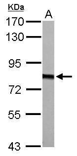
- Western Blot using HADHA Polyclonal Antibody (Product # PA5-27348). Sample (30 µg of whole cell lysate). A: Molt-4. B: Raji. 7.5% SDS PAGE. HADHA Polyclonal Antibody (Product # PA5-27348) diluted at 1:1500.

- Submitted by
- Invitrogen Antibodies (provider)
- Main image

- Experimental details
- HADHA Polyclonal Antibody detects HADHA protein by Western blot analysis. A. 50 µg mouse kidney lysate/extract.7.5 % SDS-PAGE. HADHA Polyclonal Antibody (Product # PA5-27348) dilution: 1:500.
- Submitted by
- Invitrogen Antibodies (provider)
- Main image

- Experimental details
- HADHA Polyclonal Antibody detects HADHA protein by Western blot analysis. A.50 µg rat liver lysate/extract .7.5 % SDS-PAGE. HADHA Polyclonal Antibody (Product # PA5-27348) dilution: 1:500.

Supportive validation
- Submitted by
- Invitrogen Antibodies (provider)
- Main image

- Experimental details
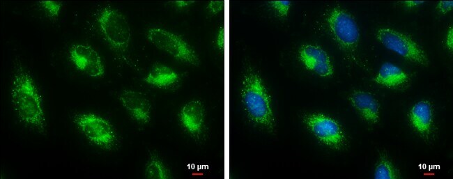
- Immunocytochemistry-Immunofluorescence analysis of HADHA was performed in HeLa cells fixed in ice-cold MeOH for 5 min. Green: HADHA Polyclonal Antibody (Product # PA5-27348) diluted at 1:500. Blue: Hoechst 33342 staining. Scale bar = 10 µm.
Supportive validation
- Submitted by
- Invitrogen Antibodies (provider)
- Main image
- Experimental details
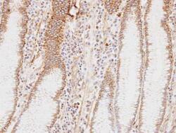
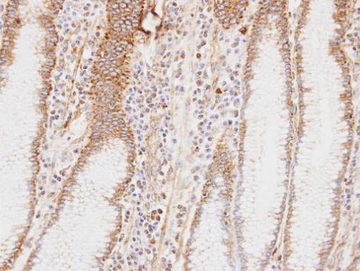
- Immunohistochemical analysis of paraffin-embedded human stomach , using HADHA (Product # PA5-27348) antibody at 1:100 dilution. Antigen Retrieval: EDTA based buffer, pH 8.0, 15 min.
Supportive validation
- Submitted by
- Invitrogen Antibodies (provider)
- Main image
- Experimental details
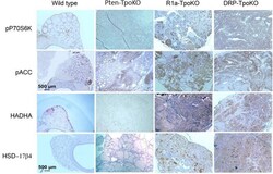
- Figure 2 Mouse tumors shown activation of mTOR and AMPK pathways. Representative mouse tumors from the genotypes shown at the top were analyzed by immunohistochemistry for the proteins shown at left. WT, Pten-TpoKO, and R1a-TpoKO mice were 1 year of age, whereas DRP-TpoKO mice were 6 months of age. All photos were taken at the same magnification, with scale bars shown in two panels in the left column.
