Explore
 Validate
 Learn
Learn Western blot
 ELISA
ELISAAntibody data
- Antibody Data
- Antigen structure
- References [3]
- Comments [0]
- Validations
- Western blot [1]
- Flow cytometry [1]
Submit
Validation data
Reference
Comment
Report error
- Product number
- ABIN648781 - Provider product page

- Provider
- antibodies-online
- Proper citation
- Antibodies-Online Cat#ABIN648781, RRID:AB_10956124
- Product name
- anti-Cysteine-Rich Secretory Protein 3 (CRISP3) antibody
- Antibody type
- Monoclonal
- Antigen
- genetic immunisation with cDNA encoding human Crisp3
- Description
- Protein G
- Reactivity
- Human
- Isotype
- IgG
- Antibody clone number
- LV-2A2
- Vial size
- 100 μg
- Concentration
- 2 mg/mL
- Storage
- short term: 2°C - 8°C, long term: -20°C
- Handling
- Avoid repeated freezing and thawing.
Submitted references Characterization and localization of cysteine-rich secretory protein 3 (CRISP-3) in the human male reproductive tract.
Cysteine-rich secretory protein-3: a potential biomarker for prostate cancer.
The human cysteine-rich secretory protein (CRISP) family. Primary structure and tissue distribution of CRISP-1, CRISP-2 and CRISP-3.
Udby L, Bjartell A, Malm J, Egesten A, Lundwall A, Cowland JB, Borregaard N, Kjeldsen L
Journal of andrology 2005 May-Jun;26(3):333-42
Journal of andrology 2005 May-Jun;26(3):333-42
Cysteine-rich secretory protein-3: a potential biomarker for prostate cancer.
Kosari F, Asmann YW, Cheville JC, Vasmatzis G
Cancer epidemiology, biomarkers & prevention : a publication of the American Association for Cancer Research, cosponsored by the American Society of Preventive Oncology 2002 Nov;11(11):1419-26
Cancer epidemiology, biomarkers & prevention : a publication of the American Association for Cancer Research, cosponsored by the American Society of Preventive Oncology 2002 Nov;11(11):1419-26
The human cysteine-rich secretory protein (CRISP) family. Primary structure and tissue distribution of CRISP-1, CRISP-2 and CRISP-3.
Krätzschmar J, Haendler B, Eberspaecher U, Roosterman D, Donner P, Schleuning WD
European journal of biochemistry / FEBS 1996 Mar 15;236(3):827-36
European journal of biochemistry / FEBS 1996 Mar 15;236(3):827-36
No comments: Submit comment
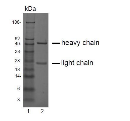
Supportive validation
- Submitted by
- antibodies-online (provider)
- Main image
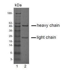
- Experimental details
- SDS-PAGE analysis of purified LV-2A2 monoclonal antibody. Lane 1: molecular weight marker, Lane 2: 2 ?g of purified LV-2A2 antibody. Proteins were separated by SDS-PAGE and stained with RAPID StainTM Reagent.
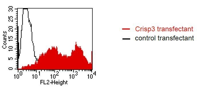
Supportive validation
- Submitted by
- antibodies-online (provider)
- Main image
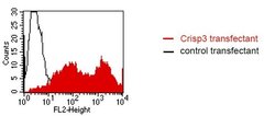
- Experimental details
- FACS analysis of BOSC23 cells using LV-2A2. BOSC23 cells were transiently transfected with an expression vector encoding either Crisp3 (red curve) or an irrelevant protein (control transfectant: black curve). Binding of LV-2A2 was detected with a PE-conjugated secondary antibody. A positive signal was obtained only with Crisp3 transfected cells.