Explore
 Validate
 Learn
Learn Western blot
 Immunocytochemistry
Antibody data
- Antibody Data
- Antigen structure
- References [0]
- Comments [0]
- Validations
- Western blot [5]
- Immunocytochemistry [1]
Submit
Validation data
Reference
Comment
Report error
- Product number
- GTX629745 - Provider product page

- Provider
- GeneTex
- Product name
- PGAM1 antibody [GT256]
- Antibody type
- Monoclonal
- Reactivity
- Human, Mouse, Rat
- Host
- Mouse
No comments: Submit comment
Enhanced validation
Supportive validation
- Submitted by
- GeneTex (provider)
- Enhanced method
- Genetic validation
- Main image
- Experimental details
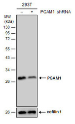
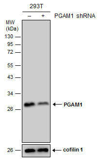
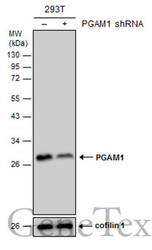
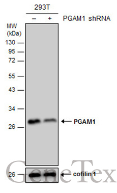
- Non-transfected (¡V) and transfected (+) 293T whole cell extracts (30 ?g) were separated by 12% SDS-PAGE, and the membrane was blotted with PGAM1 antibody [GT256] (GTX629745) diluted at 1:2000.
Supportive validation
- Submitted by
- GeneTex (provider)
- Main image
- Experimental details
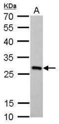
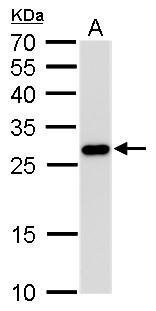
- PGAM1 antibody [GT256] detects PGAM1 protein by Western blot analysis.A. 50 ?g mouse brain lysate/extract12 % SDS-PAGEPGAM1 antibody [GT256] (GTX629745) dilution: 1:1000

- Submitted by
- GeneTex (provider)
- Main image

- Experimental details
- PGAM1 antibody [GT256] detects PGAM1 protein by Western blot analysis.A. 50 ?g Rat brain lsate/extract12 % SDS-PAGEPGAM1 antibody [GT256] (GTX629745) dilution: 1:1000
- Submitted by
- GeneTex (provider)
- Main image

- Experimental details
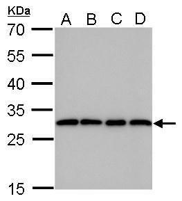
- PGAM1 antibody [GT256] detects PGAM1 protein by Western blot analysis.A. 30 ?g 293T whole cell lysate/extractB. 30 ?g A431 whole cell lysate/extractC. 30 ?g HeLa whole cell lysate/extractD. 30 ?g HepG2 whole cell lysate/extract12 % SDS-PAGEPGAM1 antibody [GT256] (GTX629745) dilution: 1:1000
- Submitted by
- GeneTex (provider)
- Main image
- Experimental details
- Non-transfected (¡V) and transfected (+) 293T whole cell extracts (30 ?g) were separated by 12% SDS-PAGE, and the membrane was blotted with PGAM1 antibody [GT256] (GTX629745) diluted at 1:2000.
Supportive validation
- Submitted by
- GeneTex (provider)
- Main image

- Experimental details
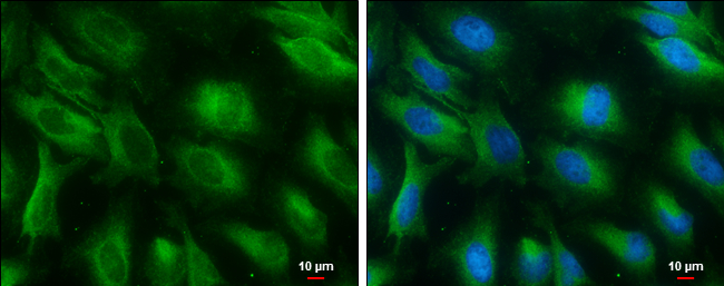
- PGAM1 antibody [GT256] detects PGAM1 protein at cytoplasm by immunofluorescent analysis.Sample: HeLa cells were fixed in 4% PFA for 15 min.Green: PGAM1 protein stained by PGAM1 antibody [GT256] (GTX629745) diluted at 1:100.Blue: Hoechst 33342 staining.Scale bar = 10 £gm.