Explore
 Validate
 Learn
Learn Western blot
 ELISA
ELISAAntibody data
- Antibody Data
- Antigen structure
- References [0]
- Comments [0]
- Validations
- Western blot [1]
- Immunocytochemistry [1]
- Flow cytometry [1]
Submit
Validation data
Reference
Comment
Report error
- Product number
- GTX31959 - Provider product page

- Provider
- GeneTex
- Product name
- PGAM1 antibody [AT1G4]
- Antibody type
- Monoclonal
- Reactivity
- Human
- Host
- Mouse
No comments: Submit comment
Supportive validation
- Submitted by
- GeneTex (provider)
- Main image
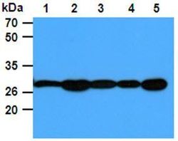
- Experimental details
- The Cell lysates (40ug) were resolved by SDS-PAGE, transferred to PVDF membrane and probed with anti-human PGAM1 antibody (1:1000). Proteins were visualized using a goat anti-mouse secondary antibody conjugated to HRP and an ECL detection system. Lane 1: 293T cell lysate Lane. 2: Jurkat cell lysate Lane. 3: Raji cell lysate Lane. 4: A431 cell lysate Lane. 5: HeLa cell lysate.

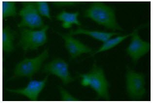
Supportive validation
- Submitted by
- GeneTex (provider)
- Main image

- Experimental details
- ICC/IF analysis of PGAM1 in HeLa cells line, stained with DAPI (Blue) for nucleus staining and monoclonal anti-human PGAM1 antibody (1:100) with goat anti-mouse IgG-Alexa fluor 488 conjugate (Green).
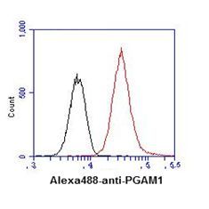
Supportive validation
- Submitted by
- GeneTex (provider)
- Main image
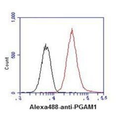
- Experimental details
- Flow cytometry analysis of PGAM1 in HeLa cell line, staining at 2-5ug for 1x10*6 cells (red line). The secondary antibody used goat anti-mouse IgG Alexa fluor 488 conjugate. Isotype control antibody was mouse IgG (black line).