 Explore
 Validate
 Learn
Learn Western blot
Antibody data
- Antibody Data
- Antigen structure
- References [2]
- Comments [0]
- Validations
- Western blot [5]
- Immunocytochemistry [2]
- Immunohistochemistry [1]
- Other assay [1]
Submit
Validation data
Reference
Comment
Report error
- Product number
- PA5-30063 - Provider product page

- Provider
- Invitrogen Antibodies
- Product name
- HSD17B3 Polyclonal Antibody
- Antibody type
- Polyclonal
- Antigen
- Recombinant protein fragment
- Description
- Recommended positive controls: Raji. Predicted reactivity: Pig (81%), Rhesus Monkey (97%), Bovine (82%). Store product as a concentrated solution. Centrifuge briefly prior to opening the vial.
- Reactivity
- Human, Mouse, Rat
- Host
- Rabbit
- Isotype
- IgG
- Vial size
- 100 µL
- Concentration
- 0.44 mg/mL
- Storage
- Store at 4°C short term. For long term storage, store at -20°C, avoiding freeze/thaw cycles.
Submitted references Regulation of uterine function during estrous cycle, anestrus phase and pregnancy by steroids in red deer (Cervus elaphus L.).
Altered expression of 3β-HSD, CYP17 and 17β-HSD in the foetal porcine gonads in response to anti-androgen flutamide exposure.
Kotlarczyk AM, Grzyb M, Korzekwa AJ
Scientific reports 2021 Oct 11;11(1):20109
Scientific reports 2021 Oct 11;11(1):20109
Altered expression of 3β-HSD, CYP17 and 17β-HSD in the foetal porcine gonads in response to anti-androgen flutamide exposure.
Knapczyk-Stwora K, Grzesiak M, Slomczynska M
Reproduction in domestic animals = Zuchthygiene 2014 Oct;49(5):725-33
Reproduction in domestic animals = Zuchthygiene 2014 Oct;49(5):725-33
No comments: Submit comment
Supportive validation
- Submitted by
- Invitrogen Antibodies (provider)
- Main image

- Experimental details
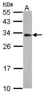
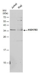
- Western blot analysis of HSD17B3 using 30 µg of Raji lysate. Samples were loaded onto a 12% SDS-PAGE gel and probed with a HSD17B3 polyclonal antibody (Product # PA5-30063) at a dilution of 1:500.
- Submitted by
- Invitrogen Antibodies (provider)
- Main image
- Experimental details
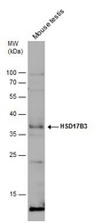
- Western Blot analysis of HSD17B3 was performed by separating 50 µg of mouse tissue extract by 12% SDS-PAGE. Proteins were transferred to a membrane and probed with a HSD17B3 Polyclonal Antibody (Product # PA5-30063) at a dilution of 1:500.
- Submitted by
- Invitrogen Antibodies (provider)
- Main image
- Experimental details
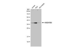
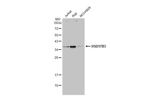
- Western Blot using HSD17B3 Polyclonal Antibody (Product # PA5-30063). Various whole cell extracts (30 µg) were separated by 12% SDS-PAGE, and the membrane was blotted with HSD17B3 Polyclonal Antibody (Product # PA5-30063) diluted at 1:1,000. The HRP-conjugated anti-rabbit IgG antibody was used to detect the primary antibody.
- Submitted by
- Invitrogen Antibodies (provider)
- Main image

- Experimental details
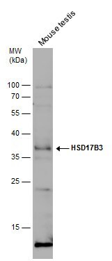
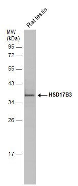
- Western Blot analysis of HSD17B3 was performed by separating 50 µg of rat tissue extract by 12% SDS-PAGE. Proteins were transferred to a membrane and probed with a HSD17B3 Polyclonal Antibody (Product # PA5-30063) at a dilution of 1:500.
- Submitted by
- Invitrogen Antibodies (provider)
- Main image
- Experimental details
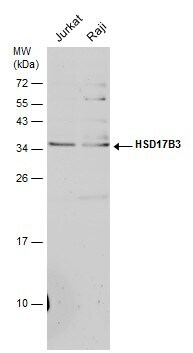
- Western Blot analysis of HSD17B3 was performed by separating 30 µg of various whole cell extracts by 12% SDS-PAGE. Proteins were transferred to a membrane and probed with a HSD17B3 Polyclonal Antibody (Product # PA5-30063) at a dilution of 1:500.
Supportive validation
- Submitted by
- Invitrogen Antibodies (provider)
- Main image
- Experimental details
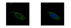
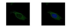
- Immunofluorescent analysis of HSD17B3 showing staining in the cytoplasm of HeLa cells. HeLa cells were fixed in ice-cold MeOH for 5 min and stained using a HSD17B3 polyclonal antibody (Product # PA5-30063) diluted at 1:500. Blue: Hoechst 33342 staining.

- Submitted by
- Invitrogen Antibodies (provider)
- Main image
- Experimental details
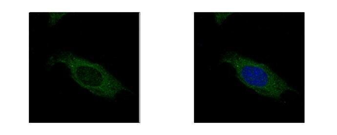
- HSD17B3 Polyclonal Antibody detects HSD17B3 protein at cytoplasm by confocal immunofluorescent analysis. Sample: HeLa cells were fixed in ice-cold MeOH for 5 min. Green: HSD17B3 protein stained by HSD17B3 Polyclonal Antibody (Product # PA5-30063) diluted at 1:500. Blue: Hoechst 33342 staining. [Images captured by Olympus FV10i Confocal Laser Scanning Microscope].
Supportive validation
- Submitted by
- Invitrogen Antibodies (provider)
- Main image

- Experimental details
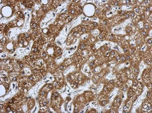
- Immunohistochemical analysis of paraffin-embedded human hepatoma, using HSD17B3 Polyclonal Antibody (Product # PA5-30063) antibody at 1:500 dilution. Antigen Retrieval: EDTA based buffer, pH 8.0, 15 min.
Supportive validation
- Submitted by
- Invitrogen Antibodies (provider)
- Main image
- Experimental details
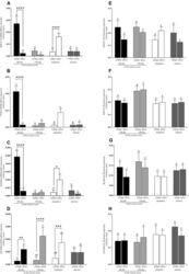
- Figure 1 mRNA and protein expression of AKR1C1 ( A , E ), P450 ( B , F ), 3beta-HSD ( C , G ) and 17beta-HSD ( D , H ) in uterine tissues (endometrium and myometrium) on 4th and 13th day of estrous cycle, in pregnancy and anestrus phase. Data were normalized against GAPDH for mRNA expression and against beta-actin (ACTB) for proteins expression. Each bar represents one experimental group with SEM. Statistical differences were analyzed by two-way analysis (ANOVA) of variance followed by the Bonferroni post hoc test using GraphPad PRISM (Version 8.3.0). The lowest statistical significance was P < 0.05. Asterisks indicate statistical differences between endometrium and myometrium (* P < 0.1; ** P < 0.01; *** P < 0.001; **** P < 0.0001). Different letters indicate statistical differences ( P < 0.05) between the experimental groups throughout endometrium (A, B) and myometrium (a-b) respectively.
